As Spinal canal is called the spinal canal. The spinal cord and the cauda equina run through it.
What is spinal canal?
At the spinal canal (Vertebral canal) is a canal that is formed by vertebral holes lying one above the other in the spine. Its course extends from the first cervical vertebra through the cervical spine (Cervical spine), the thoracic spine (thoracic spine) and the lumbar spine (LWS) to the sacrum.
The spinal cord and cauda equina pass through the vertebral canal. The vertebral canal is also called the spinal canal or spinal canal. Injuries to the spinal canal can have serious consequences. In the worst case, there is a risk of paraplegia.
Anatomy & structure
The spinal canal starts at the occipital foramen (large hole). From there it runs over the cervical spine, thoracic spine and lumbar spine in the direction of the sacrum (os sacrum).
On the abdominal side, the vertebral bodies (corpora vertebrae) and the intervertebral discs delimit the spinal canal. This is the case on the side and the back area through the vertebral arches (arcus vertebrae). In the space between two neighboring vertebrae there is an intervertebral hole (foramen intervertebrale) on both sides, which serves as an opening for the paired spiral nerves.
The spinal canal is equipped with two sturdy elongated ligaments. They are named Ligamentum flavum and Ligamentum longitudinale posterius (posterior longitudinal ligament). While the longitudinal posterior ligament is located on the front of the spinal canal, the flavum ligament is located on its back.
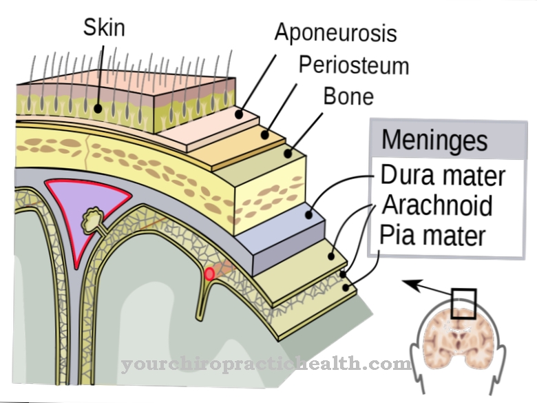
The spinal cord, which is located inside the spinal canal, is surrounded by the membranes of the spinal cord (meninges), which are special layers of tissue. The outermost layer is the periosteum, which is fused with the vertebrae. It is also called the stratum periostale or outer sheet. Under the outer leaf lies the meningeal stratum (outer membrane of the spinal cord of the dura mater spinalis). The so-called spider web skin (arachnoidea spinalis) belongs to it. This is followed by the pia mater spinalis (soft spinal cord skin).
In the spinal canal there are also several spaces between the spinal cord membranes. This includes u. a. the epidural space (Spatium epidurale), which is located between the periosteum and the stratum meningeal. The epidural venous plexus and fatty tissue are located there. The subdural space (Spatium subdurale), which is located between the arachnoid spinalis and the dura mater spinalis, forms another space. The last space is the subarachnoid space (subarachnoid space) between the pia mater spinalis and the arachnoidea spinalis. In this room there is brain water (liquor).
The blood vessels that supply the spinal cord are also found in the area of the spinal canal. The spinal cord branches (rami spinales) of the arteriae lumbales, the arteria vertrebralis and the arteriae intercostales posteriores contribute to this. A dense network of vessels is formed epidurally by the veins. This includes the ventral vertrebral plexus, which is located on the front. This area of the spinal canal is particularly vulnerable to injury if an operation is performed in its vicinity.
Function & tasks
The spinal canal houses the spinal cord, which together with the brain forms the central nervous system. The spinal cord is important for communication between the brain, internal organs, skin and muscles. At its most extensive point, the spinal cord is about the width of a finger. In adults, the spinal cord ends at the first lumbar vertebra.
However, before birth, it extends towards the sacrum. In babies, it extends to the lower lumbar vertebrae because the growth of the spine is faster than the development of the spinal cord. This phenomenon allows the spiral nerves that emerge from the spinal canal to travel a longer passage in the spinal canal in the lower section before they leave it. From the end of the spinal cord on the 1st lumbar vertebra, only the spiral nerves are then located in the spinal canal, from which the so-called horse's tail (cauda equina) is formed.
Diseases
The spinal canal can be affected by injuries or illnesses. One of the most common impairments is spinal stenosis, which leads to a narrowing of the spinal canal. Older people are particularly affected. While the lumbar and cervical spine are most likely to suffer from spinal stenosis, the thoracic spine is rarely affected.
Causes of a narrowing of the spinal canal are natural aging processes, lack of exercise, bone loss (osteoporosis) or predisposition. Sometimes several factors apply at the same time. In most cases, spinal wear and tear is responsible for spinal stenosis. The intervertebral discs, which are located between the vertebral bodies, increasingly lose fluid and height over the years. The space between the vertebral bodies is reduced and the lack of damping results in greater stress. Due to the loss of height, the ligaments lose elasticity. In some cases, the narrowing is already congenital.
Spinal canal stenosis does not always cause symptoms. Usually symptoms only develop over time. Those affected mostly suffer from muscle tension in the lower back, back pain that radiates into the leg and restricted movement in the lumbar spine. If the spinal stenosis progresses further, there is a risk of abnormal sensations such as feelings of cold, tingling, burning and sensory disorders in the legs, problems with urination or bowel movements, incontinence and disorders of sexual functions.
The most serious injuries to the spinal canal include herniated discs and vertebral fractures. If the spinal cord is injured, there is a risk of paraplegia. If the blood vessels tear, bleeding between the meninges is possible, which puts pressure on the spinal cord.

