The Masticatory muscles consists of four paired muscles that belong to the skeletal muscles and are known in medical terminology as Musculi masticatorii are designated. They move the lower jaw and enable chewing and grinding movements.
What are the masticatory muscles?
The masseter, temporalis, medial pterygoid and lateral pterygoid muscles belong to the masticatory muscles. They are present on both sides of the skull. Other muscles are involved in the process of chewing, such as various facial muscles and the muscles of the tongue and floor of the mouth, but these are not counted as masticatory muscles.
The largest muscle is the temple muscle (musculus temporalis). It arises from the temporal bone and attaches to the lower jaw. He closes the jaw and can pull it back. The masseter muscle is also involved in the closing movement of the jaw, but it also enables grinding movements. The inner (musculus pterygoideus medialis) and the outer wing muscle (musculus pterygoideus lateralis) close the jaw, enable grinding movements and, if used on one side, move the jaw to the side. All muscles of the masticatory muscles are innervated by branches of the mandibular nerve, one of the main branches of the 5th cranial nerve (trigeminal nerve).
Anatomy & structure
The masseter muscles are paired, there are four on each side of the skull. The largest and strongest is the temporal muscle. It arises from the temporal fascia (Fascia temporalis) and the temporal bone fossa (Fossa temporalis) and attaches to the crown process of the lower jaw (Processus coronoidus).
It is innervated by the deep temporal nerves (Nervi temporales profundi), a branch of the mandibular nerve. The jaw muscle is a feathered muscle and consists of a deep part (pars profunda) and a superficial part (pars superficialis). The deep part has its origin at the back third of the zygomatic arch, while the superficial part comes from the front two thirds. The attachments of the masseter muscle are the outer part of the lower jaw angle (angulus mandibulae) and a rough spot on the lower jaw, the masseteric tuberosity. The masseteric nerve, also a branch of the mandibular nerve, provides the innervation of this muscle.
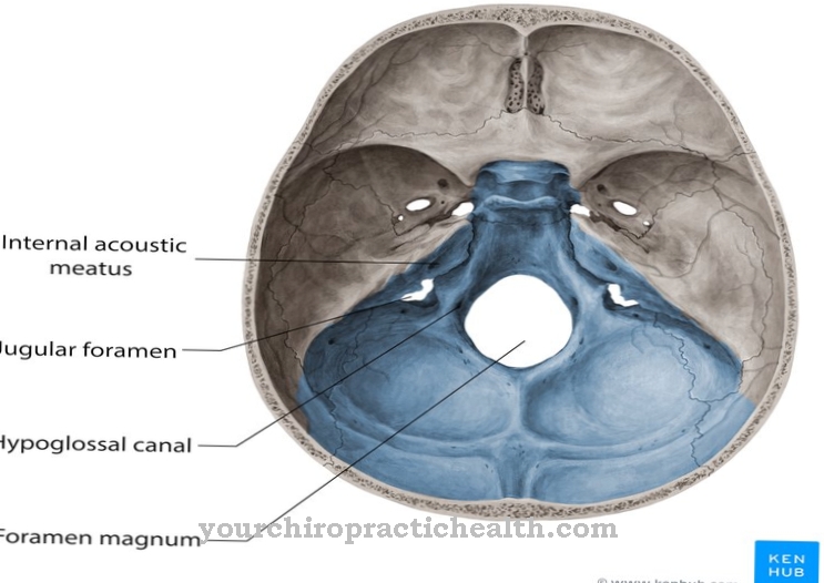
The inner wing muscle arises from a depression at the base of the skull, the pterygoid fossa, and attaches to the pterygoid tuberosity on the inner surface of the lower jaw. It is innervated by the medial pterygoid nerve. The external wing muscle is a two-headed skeletal muscle. While the upper muscle head originates from the large wing of the sphenoid bone (Ala major), the lower head originates from a bone process of the sphenoid bone, the pterygoid process. The outer wing muscle is innervated by the lateral pterygoid nerve.
Function & tasks
The very strong temporal muscle takes on almost 50% of the force that is necessary for the chewing movement. He can close the jaw (adduction of the jaw) as well as push it forward (protrusion) and withdraw it (retrusion). The vertical muscle fibers are mainly used for adduction, while the horizontal fibers are primarily used for pro- and retrusion.
If the temporal muscle is only used on one side, the lower jaw is shifted to the side (laterotrusion). The masseter muscle is also involved in jaw closure. He also lifts the lower jaw and can pull it forward. This muscle also helps maintain the tension in the temporomandibular joint capsule. The inner wing muscle supports the masseter muscle in closing the jaw. But since it is narrower, it can only muster half as much force. When it contracts, the jaw not only closes, but also moves forward.
With a unilateral contraction, he shifts the lower jaw to the side, that is, he makes grinding movements possible. The outer wing muscle has a special position among the jaw muscles because it initiates the opening of the mouth. This movement is taken over and continued by the suprahyoid muscles of the floor of the mouth. This muscle is also involved in advancing the jaw and in grinding movements.
You can find your medication here
➔ Toothache medicationDiseases
Common complaints are pain when chewing or cracking and crunching noises. They are mostly caused by tense mastication muscles. This tension can either be caused by strong active tension, such as in states of anxiety or fits of anger, or they arise from bad bites.
If the bite is in the correct position, the temporomandibular joints, bones and muscles work in harmony with one another, while a bad bite can lead to uneven loading and thus excessive tension in the masticatory muscles. Nocturnal crunching or lengthy dental interventions can also cause painful muscular tension. Often the pain spreads further and radiates into the teeth or into the head, whereby the cause is wrongly suspected to be somewhere other than the muscles. The pain in the masticatory muscles is known as craniomandibular dysfunction (CMD) or temporomandibular disorders (TMD).
Treatment is based on the cause. If there is a wrong bite, this will be corrected as far as possible. To prevent crunching at night, the dentist adjusts a so-called grinding splint, which is intended to prevent teeth from rubbing against each other. The jaw clamp represents a further disorder in the area of the jaw muscles. It is no longer possible to open the mouth due to severe muscle spasms. This spasm of the chewing muscles is also known as trismus.
A distinction is made between different degrees, which are based on the distance between the tooth edges of the front teeth of the upper and lower jaw. With grade I the restriction of the opening is only minimal, with grade II the distance between the tooth edges is about 10 mm and grade III only allows an opening of 1 mm.





.jpg)

.jpg)

.jpg)
