The Ala minor ossis sphenoidalis is part of the human skull. They are near the sphenoid bone. Your job is to help shape the eye socket.
What is the ala minor ossis sphenoidalis?
The ala minor ossis sphenoidalis belong to the human skeletal system. They are translated as small sphenoid wings. The sphenoid bone and the wing that goes with it are components of the skull.
They are made of a bony structure. They are located in the back of the face. The sphenoid bone forms the back of the human eye socket. The ala minor ossis sphenoidalis consist of thin plates of bone. They are divided into the anterior wing of the sphenoid bone and the posterior wing of the sphenoid bone. Both are laid out on both sides and optically have a wing shape. They are formed from triangular bone plates.
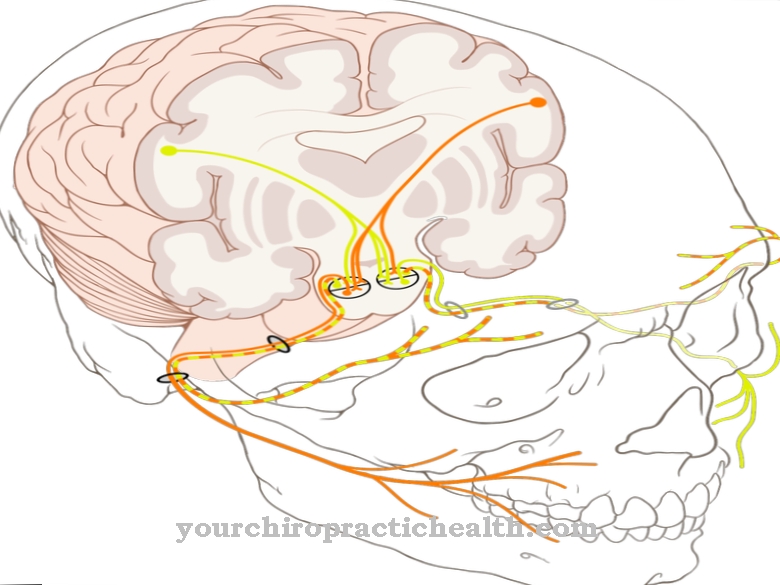
The wings of the sphenoid bone form small openings and thus passageways for nerves and vessels. The wings of the sphenoid bone are very small parts of the human skull. They serve as the exit point for the optic nerve or the V cranial nerve. The eye can be supplied through the openings of the ala minor ossis sphenoidalis and the recorded visual information is transported via the nerve pathways to the brain for further processing.
Anatomy & structure
The ala minor ossis sphenoidalis is a collective term for various small bones of the sphenoid wing. are divided into the wings of the anterior sphenoid and the posterior sphenoid. The bilateral wings of the anterior sphenoid bone are called the ala ossis praesphenoidalis or ala minor.
They form part of the back of the eye socket, the orbit. Both front wings of the sphenoid bone are traversed by the optic canal. The optic canal consists of the optic nerve, the optic nerve and the ophthalmic artery. The wings of the posterior sphenoid are also placed on both sides. They are known as Ala ossis basisphenoidalis or Ala major. The foramen ovale is located in them. This serves as the exit point for the V cranial nerve, the mandibular nerve.
At the posterior end of the wing of the sphenoid bones is the foramen spinosum. This serves the arteria meningea media when it enters the cranial cavity. The ala minor ossis sphenoidalis have a triangular and thus wing-shaped shape. Since they form the rear area of the eye socket, they can only be felt to a small extent from the outside in the area of the temples.
Function & tasks
The task of the bone plates of the ala minor ossis sphenoidalis is to shape the posterior eye socket. The human eye is protected in a cave. This is completely surrounded by different structures of the brain skull. Light and colors are absorbed through the eye. All visual stimuli are picked up by the individual components of the eye and transported to the cortex via the optic nerve. There they are evaluated and reactions initiated.
The optic nerve runs through the ala minor ossis sphenoidalis. This ensures that, on the one hand, the eye is supplied and, at the same time, recorded stimuli reach the visual cortex as quickly as possible. This is in the back of the human head. Some visual stimuli are processed within a few milliseconds. For this to happen, the optic nerve needs a fast path to process stimuli.
In addition, various blood vessels pass through the ala minor ossis sphenoidalis. The ophthalmic artery is one of them. The arterial blood vessel supplies the eye and the eye socket with important messenger and nutrients. The branches of the ophthalmic artery supply the retina, the lacrimal gland, the lens and also the ethmoid cells. The V cranial nerve, the mandibular nerve, also runs through the Ala minor ossis sphenoidalis. The cranial nerve supplies large areas of the face. With its ramifications, for example, the teeth, the jaw, the cheek, the auricle, the palate or the chin are supplied.
You can find your medication here
➔ Medicines for eye infectionsDiseases
Lesions of the skull are mostly caused by severe impacts. This happens as a result of falls or accidents. The bones of the human skull are very stable and can withstand minor damage mostly without further consequences.
A lesion on the bone can cause a break or bruise. In most cases, the damage heals on its own after a few weeks. Bruises are considered uncomfortable and cause headaches.The patient should take it easy and avoid physical exertion or pressure on the head during the healing process. The bones of the posterior socket are rarely damaged on their own. Usually the adjacent regions are affected, so that the wing of the sphenoid bone is to be ascribed to another cause as an accompanying symptom. If tissue swelling occurs inside the skull, it can lead to the openings of the ala minor ossis sphenoidalis becoming blocked.
The nerve tracts and vessels can then no longer be used as passage points. On the one hand, this means that blood circulation can become blocked. If they are not corrected or if the blood cannot find another way, bleeding is the result. These can cause impaired consciousness, loss of consciousness, or headaches. In addition, sensorimotor failures usually occur.
There is a risk of a stroke. In addition, the nerve tracts can no longer adequately supply the organs. As soon as the openings of the Ala minor ossis sphenoidalis are closed, the eye is no longer adequately innervated. This leads to loss of vision. Recorded information can no longer be transported into the visual cortex and the function of the rods and cones in the eye is restricted.

