As Aponeuroses are mostly flat tendon plates made of connective tissue, which are used for the sinewy attachment of muscles. In addition to the hand, foot and kneecap, the abdomen, the palate and the tongue have aponeuroses. The most common disease of the tendon plates is inflammation known as fasciitis.
What is an aponeurosis?
The medical term aponeurosis comes from Latin. Literally translated the term means something like Tendon plate. This refers to the flat or flat connective tissue structures that serve the tendinous attachment of one or more muscles and appear as an extension of muscle tendons.
Well-known examples of aponeuroses are, besides the palate aponeurosis, the palmar aponeurosis, the plantar aponeurosis, the rectus sheath, the tongue aponeurosis and the retinaculum patellae. The plantar fascia tightens the arch of the foot and maintains it. It protects the muscles, nerves, blood vessels and tendons on the soles of the feet. The palmar fascia on the hand has similar functions. The structure of the aponeuroses differs with the localization. The aponeurosis differs from other types of connective tissue mainly in its function and its anatomically layer-like shape. All aponeuroses are always directly related to at least one muscle and its tendon.
Anatomy & structure
The palatal aponeurosis is a fiber-rich layer of connective tissue that serves as the basis for the soft palate. Palate muscles to move the palate radiate into the connective tissue.
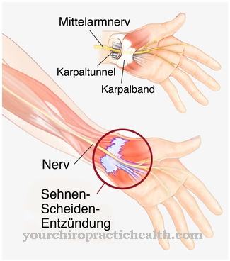
The palmar aponeurosis consists of complex, three-dimensionally arranged longitudinal, transverse and vertical fibers and is connected to the surface fascia of the hand by fibrous connective tissue. It lies in the central palm on the short palm muscles and fuses laterally with the fasciae of the hypothenar and thenar muscles. The plantar fascia has roots in the calcaneus and diverges in a V-shape in the metatarsophalangeal joint capsules and the flexor tendons of the metacarpal joint.
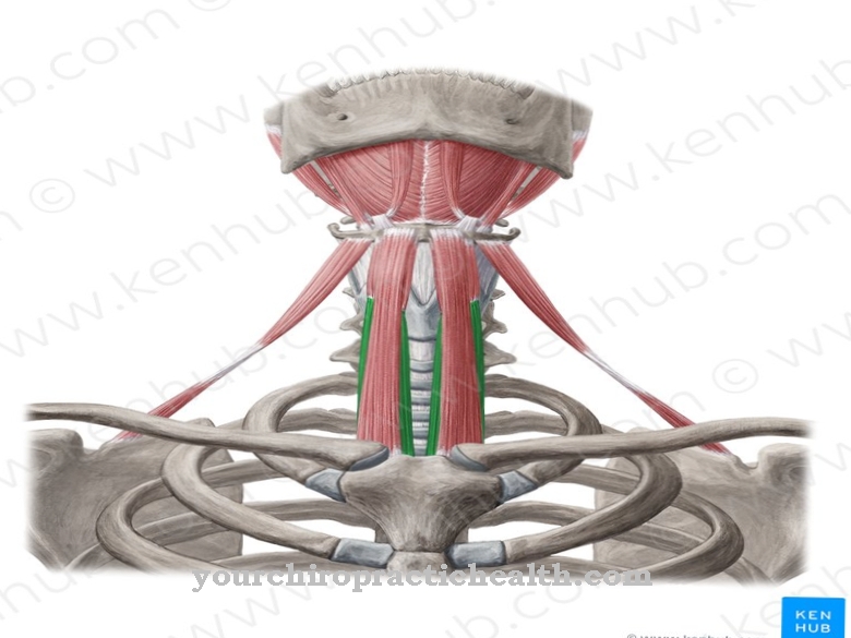
The rectus sheath consists of the aponeuroses of the three abdominal wall muscles, Musculus obliquus internus abdominis, Musculus transversus abdominis and Musculus obliquus externus abdominis. It envelops the rectus abdominis muscle. The tongue aponeuros is a tough layer of connective tissue between the lining of the tongue and the tongue muscles. The aponeurosis retinaculum patellae supports the kneecap and is part of the outer joint capsule layer of the knee joint.
Function & tasks
The main task of all aponeuroses is the formation of the muscle tendon attachment. In this context, the palatal aponeurosis is often referred to as the functional tendon lengthening of the tensor veli palatini muscles. According to current knowledge, this aponeurosis is more of an extension of the adjacent bone periosteum. The palmar aponeurosis is irreplaceable for the grasping movement of the hand. It tightens the skin on the palmar side of the hand.
Because of its fiber lines, it creates close contact between the gripped object and the hand and at the same time protects the blood vessels and nerves under the connective tissue layer. The plantar fascia stabilizes the longitudinal arch of the foot skeleton. It has an ideally functional lever arm for bracing the arch of the foot. The aponeurosis is fused into the skin of the sole of the foot through dense fiber bundles and fixes the skin through this tight anchoring. This creates the basis for a secure stand. The fat pads between the fiber strands serve as pressure pads. The muscle fibers of the abdominal wall are shortened by the rectus sheath. If the abdominal wall contracted too tightly, the abdominal cavity would be narrowed and the organs would not have enough space.
The rectus sheath also closes the tendon plates of the abdominal muscles into one unit. The tongue aponeurosis serves as a stable attachment for the tongue muscles and the retinaculum patellae forms a retaining strap for the kneecap. Accordingly, all aponeuroses have a stabilizing and holding function in common. Usually the connective tissue layers also take on protective functions. Despite these tasks, the structures are rather passive structural elements.
You can find your medication here
➔ Medicines for muscle weaknessDiseases
Every aponeurosis in the body can be affected by inflammation. This pathological phenomenon is also known as fasciitis and most often affects the plantar fascia on the foot. If the plantar tendon plate is inflamed, the doctor speaks of plantar fasciitis.
Usually this phenomenon is preceded by overuse of the associated muscles. Such overloads occur mainly when doing sports, jumping or running. Dancing, soccer, and basketball are all considered risk factors. In addition to overload, the inflammation can also be caused by previous injuries to the foot. Plantar fasciitis manifests itself as severe pain in the heel area, which usually increases with exercise. The beginning is creeping. As the disease progresses, symptoms worsen over weeks or even months. The pain can cause inability to walk on the climax of the disease. Usually the pain shoots in strongly at the beginning of a load, but disappears within a certain load duration.
The foot aponeuroses also affects Ledderhose's disease, which causes a thickening of the connective tissue and corresponds to a fibromatosis. On the hand aponeurosis, the same phenomenon is called Dupuytren's disease. With both phenomena, nodes form in the aponeuroses that slowly increase in size. Painful lumps can limit mobility. Therefore, although both conditions are considered benign conditions, surgical removal may be indicated.
The primary cause of the growths is so far unknown. The myofibroblasts cause the connective tissue to multiply. Current research is concerned with the factors that stimulate it. It has been speculated that injuries, genetic components, primary diseases such as diabetes mellitus, and nicotine or alcohol consumption could all play a role in the disease's etiology. For all patients with a benign connective tissue overgrowth in a certain part of the body, there is an increased risk of suffering further connective tissue overgrowths.









.jpg)
