As a branch of the facial artery supplies the Angular artery the eye ring muscle, the lacrimal sac and the orbital and infraorbital regions. Arterial damage, for example from an aneurysm and / or an embolism, can lead to necrosis of the affected tissue.
What is the angular artery?
The angular artery represents a branch of the facial artery (arteria facialis or arteria maxillaris externa). The facial artery supplies numerous regions of the head with blood, in which it transports oxygen, among other things.
Since the angular artery is part of the body's circulation, it carries the vital breathing gas from the lungs to the tear sac, the eye ring muscle and the skin in the orbital and infraorbital regions. Regardless of where they are, cells that suffer from a lack of oxygen and energy can no longer adequately carry out their tasks and die after a while if the deficiency persists. In such a case, the affected tissue becomes necrotic.
Anatomy & structure
After various branches have branched off from the facial artery, the angular artery remains as the terminal branch of it. It runs from bottom to top along the nose and finally leads to the inner corner of the eye. Its course is similar to that of the angular vein.
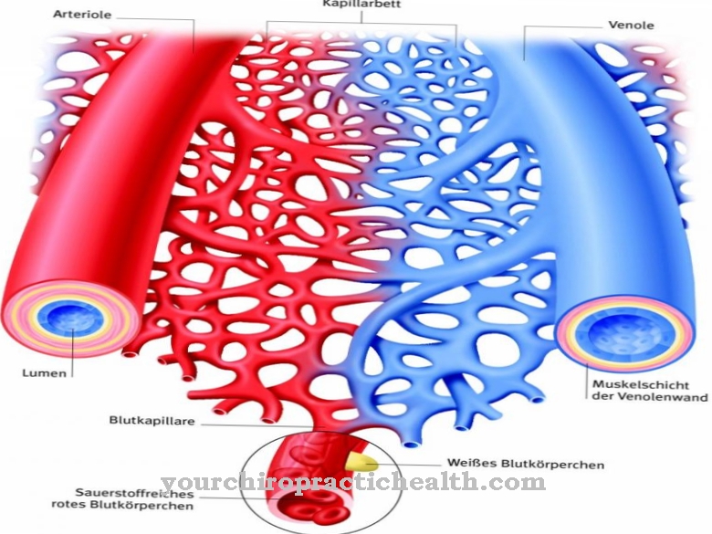
In contrast to the angular vein, the angular artery transports oxygen-rich blood. The red blood cells (erythrocytes) are loaded with it in the lungs and initially flow to the heart. The powerful muscle then pumps the blood into the main artery (aorta), from where the blood takes two different routes depending on the side of the body. On the left, the aorta directly feeds the common carotid artery (common carotid artery).
In the right half of the body, the blood flows from the aorta into the arm and head vascular trunk (Truncus brachiocephalicus), which is also known as the Arteria innominata or Arteria anonyma and whose branches on the right side include the common carotid artery. The common carotid artery then divides into the inner and outer carotid arteries (internal carotid arteries and external carotid arteries). The arteria facialis branches off from the latter and finally merges into the arteria angularis.
Natural connections to other blood vessels exist between the arteria angularis and the arteria infraorbitalis as well as the arteria dorsalis nasi. The anatomy calls such connections anastomoses.
Function & tasks
The tasks of the angular artery include supplying the tear sac. This is located in the inner corner of the eye in the lacrimal sac (fossa lacrimalis) and belongs to the lacrimal apparatus (apparatus lacrimalis). In addition, the angular artery is responsible for the blood supply to the eye ring muscle (orbicularis oculi muscle).
This lies in the eye socket around the organ of vision. A contraction of the orbicularis oculi muscle closes the eyelid and helps squint the eyes. In addition, the eye ring muscle is able to widen the tear sac, which makes it easier for the tear fluid to drain away. It finally reaches the nose through the nasal duct (ductus nasolacrimalis), which is 20 to 25 mm long. The facial nerve (Nervus facialis) is responsible for the neuronal innervation of the muscle orbicularis oculi; its motor fibers control the tension and relaxation of the muscle.
The angular artery also supplies oxygen-rich blood to two areas of the skin. The orbital region is located on the eye and also includes the eyelids and the eye itself. Below the orbital region is the infraorbital region, the cells of which are also dependent on the blood supply to the angular artery.
Diseases
An aneurysm of the angular artery can cause the death of the skin cells that lie in the affected area if the tissue becomes undersupplied as a result. Aneurysms can manifest not only on arteries, but also on the heart chambers and veins.
The affected blood vessel sags when an aneurysm develops, thereby forming a pouch. The extension can also take on a spindle-shaped shape. The aneurysm greatly expands the wall of the affected artery and reduces its flexibility. This can rupture the blood vessel. In addition, blood clots (thrombi) may be deposited in the aneurysm. When they loosen, they can create a blockage in narrower parts of the vein, which medicine calls an embolism. In addition to thrombi, fat, lime, undissolved gases and foreign bodies can also cause such an occlusion.
The skin regions that the angular artery is responsible for supplying can necrotize as a result of damage from an aneurysm. At first glance, the clinical appearance resembles damage that is possible in some cases through the use of so-called dermal fillers in plastic surgery.
However, as part of such cosmetic interventions, in some cases the arteries of the face, which are located in the corresponding region, are also damaged. You may experience redness, blistering, and other skin changes; sharp pain may indicate embolism of the artery. During treatment, doctors may need to remove the necrotized areas of skin.
Angular artery embolism is also possible when a thrombus forms in another blood vessel and flows to it through the bloodstream. However, vascular occlusion caused by such thrombi affects the angular artery less often than the arteries that flow to the brain. Since the angular artery anastomises with the nasal dorsal artery, the effects can also show up in areas for which the nasal dorsal artery is responsible. This includes the skin of the bridge of the nose and the bridge of the nose.



.jpg)

.jpg)
.jpg)


.jpg)