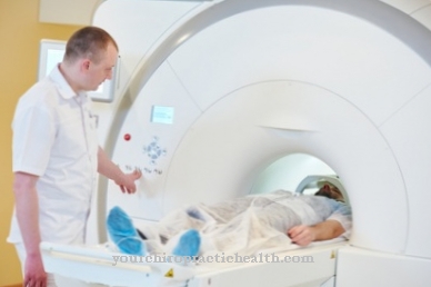
The Arthrography is an invasive imaging method in radiology that depicts the soft tissue structures of joints using double contrast medium administration. The diagnostic and differential diagnostic method is therefore particularly relevant with regard to inflammatory and degenerative joint diseases. In the meantime, MRT and CT have largely replaced arthrography, but arthrography is still used to examine the shoulder joint, regardless of these two newer and even more precise imaging methods.
What is the arthrography?

Arthrography is an imaging examination method used in radiology. It is primarily of diagnostic and differential diagnostic importance. In the invasive procedure, the radiologist examines the joints and depicts their bony structures, including all soft tissue structures, using X-ray imaging.
The soft tissue structures include, above all, the cartilaginous joint coatings on the joint surfaces, the joint discs and the joint fluid. The joint chambers, the tendon sheaths and the bursa are also shown in the pictures. These structures are displayed by means of intravenous contrast agent administration, which allows all fine structures to emerge in the imaging. The soft tissue structures shown in this way would not be visible on a conventional X-ray, but they could be seen on MRT or CT images. For this reason, with the increasing popularity of MRI and CT, arthrography has now almost survived.
Function, effect & goals
In arthrography, various joint interiors are shown with their individual structures. This makes the procedure particularly relevant in relation to inflammatory joint diseases such as arthritis or degenerative joint diseases such as osteoarthritis. However, malformations such as so-called hip dysplasia can also be visualized in the procedure. Even traumatic and tumorous joint diseases can be visualized using arthrography. Ultimately, all joints of the body can be represented using the method.
However, this type of imaging is currently most common in the shoulder joint. In this context, the imaging can show a dislocated shoulder, for example. The procedure is also indicated in the case of impingement syndrome, i.e. when the shoulder is overloaded by physical activity. In the case of impingment syndrome, for example, arthrography shows a thickened and pinched supraspinatus tendon, which has a detrimental effect on the shoulder joint. Arthrography can also be used to diagnose a rupture of the shoulder joint muscles. In addition to the shoulder joint, joints such as the elbow joint, the wrist and the hip joint as well as the knee joint, the ankle joint or the finger joints can also be shown. In most cases, however, the examination is not necessary for these joint connections, since MRI or CT can serve the same purpose.
In order to have an arthrography carried out, the patient turns to an appropriately equipped radiology department. The radiology staff pays strict attention to sterile conditions during the examination. For example, the patient's skin is carefully disinfected beforehand. The attending physician then punctures the joint space. Usually under fluoroscopy, he injects the contrast agent into it. In addition to the positive X-ray contrast medium, negative air is usually also used as a contrast medium in arthrography, as is common in pneumarthrography, for example. This double contrast procedure shows the joint most precisely. After the administration of the contrast medium, recordings are made in two planes and medically assessed.
Risks, side effects & dangers
Before MRI, CT and sonographic imaging were available, arthography was the only option for soft tissue imaging. That has changed in the meantime and arthrography is therefore no longer justified as a method. Today, MRI or sonography imaging are more likely to be used for the same purpose. MRI in particular depicts soft tissues in joints even more precisely.
On the other hand, arthography for complaints in the carpal and shoulder joints is still a standard procedure that is conventionally combined with magnetic resonance imaging or CT. In addition, both X-ray and MRT and CT procedures are, in a certain sense, arthographies that are now implemented via the administration of contrast media. In the X-ray image, air is used as a contrast medium to show the soft tissues. In MRI you work with a water-soluble contrast agent and in CT, air and water-soluble contrast agent are used in combination.
The fact that actual arthrography is now rarely used is not least due to the risks of the inversive procedure. As a rule, the patient tolerates the procedure well, nonetheless there may be side effects. A professional staff is the top requirement for an arthrography, since under non-sterile conditions, for example, severe inflammations and infections can occur. Because the joint is punctured during the procedure using contrast agent, this partial step can also cause pain. With professional, experienced staff this risk of pain is reduced. In the past, the administration of contrast media itself was associated with considerable risks, as some carcinogenic agents were used.
Today, water-soluble contrast media are usually either iodine- or gadolinium-based, which limits their harmful effects.Nevertheless, as a contraindication, allergic reactions to iodine or gadolinium can occur in rare cases. Apart from that, the administration of contrast medium can cause nausea or headaches. Sports activities should not be undertaken on the same day. Before the examination, the patient takes part in an extensive consultation, which informs him of all risks and side effects. At the end of the interview, he signs a declaration of consent. In the case of acute inflammation, allergies to contrast media and infections, the procedure is generally not recommended.
Typical & common joint diseases
- arthrosis
- Joint inflammation
- Joint pain
- Joint swelling
- Rheumatoid arthritis


.jpg)

.jpg)
.jpg)


.jpg)