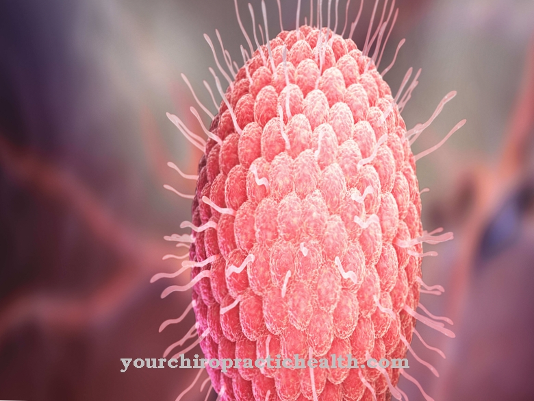
Corynebacteria are gram-positive, rod-shaped bacteria.They are immobile and grow under both aerobic and anaerobic conditions. One of their types is responsible for diphtheria, among other things.
What are Corynebacteria?
Corynebaceries are a genus of gram-positive, rod-shaped bacteria that can grow facultatively anaerobically, that is, they can exist in the presence of oxygen as well as in the absence thereof. Their species are immobile and do not form spores. They are also catalase positive and oxidase negative. In addition, the corynebacteria only grow under demanding conditions, namely at 37 ° C and the presence of 5% CO2.
Corynebacteria have a great diversity of species. Some species are pathogenic to humans (such as C. diphtheriae), other species are saprophytes, that is, they live on dying plant remains. Even more are non-pathogenic species that occur in the normal flora on the skins and mucous membranes of humans.
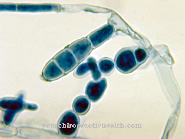
Characteristic of the Corynebacteria is the club-shaped swelling at one end, from which they got their name (Greek koryne = club). Another specialty of the corynebacteria is the presence of mycolic acids in the cell wall, which is also found in mycobacteria.
Occurrence, Distribution & Properties
Non-pathogenic types of corynebacteria occur mainly on the normal flora of the skin and the mucous membrane of humans. However, pathogenic species are also widespread and can be found worldwide. The most common infectious disease caused by a Corynebacterium is diphtheria. The transmission takes place exclusively from person to person and can take place through droplet or smear infection.
If a person is infected with a Corynebacterium, local pathogen colonization follows after an initial infection. The pathogen can then spread, or in the case of C. diphtheriae, for example, an exotoxin is formed which inhibits protein synthesis. The incubation period ranges from 2 to 10 days. In general, corynebacteria are rarely the cause of a disease, especially since there is good vaccination protection in Germany. Exceptions are diphtheria, which is endemic to Russia, and Corynebacterium minutissimum.
Corynebacteria are gram-positive rod bacteria. They have a certain pleomorphism, which means they are able to change their shape depending on the conditions of the environment. They contain mycolic acid in their cell wall and are catalase-positive, but oxidase-negative. Corynebacteria can be stained using Neisser staining and show yellow-brown bacteria with black-blue polar bodies.
Meaning & function
There are numerous types of corynebacteria that are found in the normal flora of the skin and mucous membranes. These include C. minutissimum, C. xerosis, C. pseudotuberculosis, C. jeikeium, C. pseudodiphteriticum and Corynebacterium bovis. Some species are referred to as facultative pathogenic because under certain conditions they can trigger diseases, for example a weakening of the immune system.
These species include the C. minutissimum, which causes erythrasms, and the C. jeikeium, a possible cause of sepsis. The physiologically present corynebacteria break down the fats that are secreted by the sebum glands into fatty acids. These are then responsible for the acidic environment of the skin and mucous membranes, which forms part of the protective acid mantle. This is a weakly acidic pH value that is located on the epidermis and thus has a bactericidal effect on pathogens, which leads to an inhibition of the growth of germs. The corynebacteria thus form part of the innate, unspecific immune defense. In addition, the C. striatum is said to be partly responsible for the typical armpit odor.
You can find your medication here
➔ Medication for shortness of breath and lung problemsIllnesses & ailments
The corynebacteria describe a genus of bacteria that is characterized by many species. The most important pathogenic species is C. diphtheriae. It is the causative agent of diphtheria. Humans are the only hosts of this bacterium and usually transmit the pathogen by droplet infection. C. diphtheriae then often enters the pharynx, less often in skin wounds, and multiplies there. After reproduction, it produces the diphtheria toxin, which comes from the bacteriophage. The bacteriophages are viruses that attack bacteria.
Diphtheria toxin works by inhibiting protein synthesis. A dose of 100-150 ng per kg body weight is enough to kill a person. First, there is a local effect in the throat of the person affected. The epithelial cells of the mucous membrane are destroyed, bleeding and fibrin secretions occur. The latter form the characteristic fibrin coatings on the infected mucous membrane, which is known as a pseudomembrane. Other bacteria as well as cells and blood cells get caught in the pseudomembrane.
The classic pharyngeal-larynx diphtheria is also characterized by fever, swelling of the lymph nodes and palsy of the soft palate. Dreaded complications are myocarditis, nerve and kidney damage if the toxin spreads systemically.
In the past, so-called diphtheric laryngitis was a dreaded complication that quickly led to death from suffocation. It was characterized by a Caesar's neck (severe swelling of the lymph nodes) and a sweetish halitosis. In addition to C. diphtheriae, other related species can also trigger diphtheria, for example C. ulcerans, which can also affect animals.
C. jeikeium is facultatively pathogenic and can cause sepsis. In addition, C. minutissimum can trigger erythrasma, a superficial, reddening dermatitis.










.jpg)


.jpg)

.jpg)
