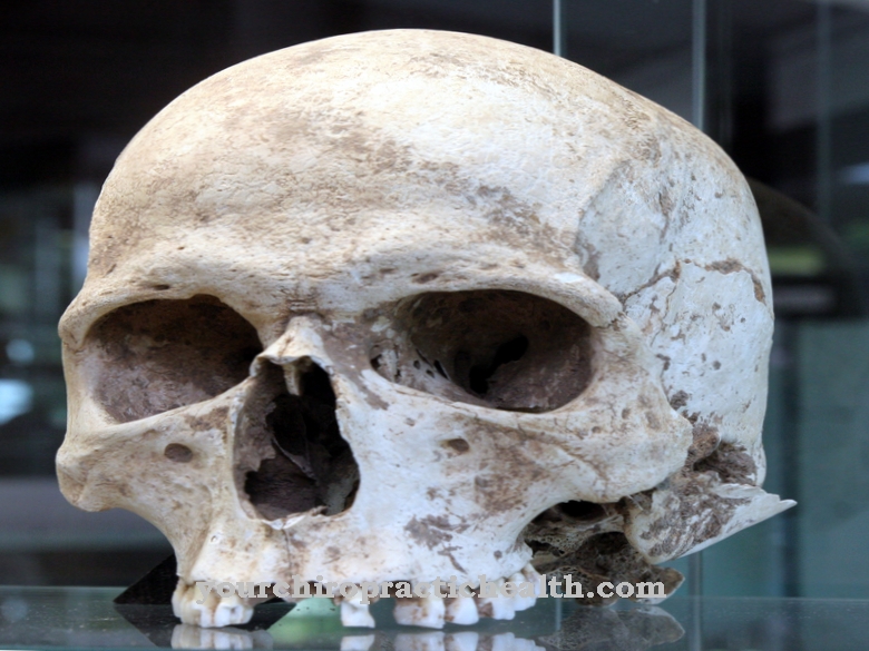
The Fossa cranii media is the middle cranial fossa, which contains the temple or temporal lobe of the cerebrum. Their shape is reminiscent of the shape of a butterfly. The middle cranial fossa also has several openings through which cranial nerves and blood vessels have access to the brain.
What is the media cranial fossa?
The human brain lies in a bony cranial cavity that provides protection and a dimensionally stable shell for the sensitive organ. The middle cranial fossa corresponds to the middle cranial fossa. It is located between the anterior cranial fossa, which lies under the frontal lobe of the brain, and the posterior cranial fossa, which is the rearmost of the three cranial pits.
All three belong to the base of the skull (base cranii), which together with the roof of the skull (calvaria) forms the brain skull. Viewed from above, the shape of the middle cranial fossa is reminiscent of a butterfly, which is symmetrically mirrored along the longitudinal axis of the skull. The middle cranial fossa supports the temporal lobe of the cerebrum (lobus temporalis). Its coils (gyri) and folds (sulci) depict the skull bones as Impressiones digitatae and Juga cerebralia.
Anatomy & structure
At the border between the middle and anterior cranial fossa is the small wing of the sphenoid bone (Ala minor ossis sphenoidalis), which encloses the fossa cranii media in a convex arch.
In the posterior area, the middle cranial fossa ends at an edge of the temporal bone (pars petrosa ossis temporalis). The "floor" of the fossa cranii media consists of several cranial bones: the large wing of the sphenoid bone (Ala major ossis sphenoidalis), the parietal bone (Os parietale), the temporal bone scale (Pars squamosa ossis temporalis or Squamosa temporalis) and the surface of the temporal bone.
There are several openings in and between the bones. These include the upper orbital fissure (fissura orbitalis superior), which creates a connection to the eye socket (orbit). The optic canal (Canalis opticus), which is 5–10 mm long, also leads to the orbit. With a size of 20 x 6 mm, the opening is relatively large. The foramen oval in the sphenoid bone has a uniformly rounded shape and is slightly smaller at 4–5 x 7–8 mm. The foramen lacerum, on the other hand, has uneven edges and lies between the sphenoid, temporal and occipital bone. The foramen spinosum and the foramen rotundum represent further penetration points in the fossa cranii media and have a round shape.
Function & tasks
The task of the middle cranial fossa is to provide protection to the part of the brain that is above it. Part of the temporal lobe is represented by the hippocampus, which plays an important role in memory. Other structures in the temporal lobe such as the entorhinal cortex as well as the parahippocampal and perirhinal areas are also decisive for the ability to remember. The Wernicke Center is part of the language center and is used for language understanding. It corresponds to the Brodman area A 22.
In addition, the temporal lobe houses the primary auditory cortex, which processes acoustic perceptions and releases nerve fibers to the internal capsule. The so-called neocortical associative areas in the temporal lobe deal with complex auditory, but also visual information. Parts of the temporal lobe can also be combined to form the limbic system. It is a system of different brain structures that are involved in the development of emotions, memory functions and sexual functions, among other things.
The limbic system is historically considered to be very old. It includes the hippocampus, the amygdala (corpus amygdaloideum or almond nucleus), the mammillary body (corpus mamillare), the cingulate gyrus and the parahippocampal gyrus. These anatomical units are closely connected to one another via nerve tracts. The activity of the amygdala is particularly important for the emotion fear and for conditional learning.
You can find your medication here
➔ Medicines against memory disorders and forgetfulnessDiseases
Various cranial nerves and blood vessels pass through the openings in the middle cranial fossa.Lesions in these areas can therefore lead to the failure of certain nervous functions. Bleeding can also potentially damage tissues.
In addition, they can lead to supply disruptions. Lesions on cranial nerves are also possible from injuries, inflammation, and tumors. A nasopharyngeal carcinoma, for example, can spread through the foramen lacerum to the cavernous sinus, which drains venous blood from the brain. There the cancer is able to damage cranial nerves in some cases. As part of the diagnosis of nasopharyngeal carcinoma, doctors often also check the function of cranial nerves III, V, VI, IX and X.
The temporal lobe of the cerebrum is located in the middle cranial fossa. In temporal lobe epilepsy, people suffer from seizures, most of which begin between the ages of 5 and 10. In temporal lobe epilepsy, medicine differentiates between a lateral / neocortical variant on the one hand and a mesial form on the other.
The entorhinal cortex, which is affected by nerve loss in the context of Alzheimer's dementia, also lies in the temporal lobe. Damage to the temporal lobe or the surgical removal of tissue can lead to memory disorders in other contexts as well. An example of this is anterograde amnesia, in which those affected can only acquire new declarative knowledge, episodic memories and other memory contents to a limited extent. The disease became famous through Henry Gustav Molaison, from whom a surgeon removed large parts of the temporal lobe in order to treat his epilepsy. As "patient H. M." his severe memory impairment caused a sensation and was extensively examined.
The causes of Wernicke's aphasia also lie in the temporal lobe. The speech disorder manifests itself as impairment of speech understanding and is also known as sensory aphasia. With bilateral temporal lobe syndrome or Klüver-Bucy syndrome, patients only show a limited ability to perceive emotions. Increased sexual behavior (hypersexuality) is possible. In addition, other symptoms such as abnormalities in visual processing can occur.



.jpg)


.jpg)


.jpg)




.jpg)
