What is the lens?
In the human eye, the one that is convexly curved on both sides is used lens to bundle incident light so that on the back of the vitreous body on the retina at the point of greatest resolution (point of sharpest vision, Fovea centralis) a sharp image is created. This is picked up by the color photosensors (mainly M and L cones for green and red) and forwarded to the visual center.By pulling the zonular fibers at the edge of the capsule, the lens can be “pulled flat” and thus accommodate to distant vision. When the tension of the zonular fibers subsides, the lens resumes its natural, almost spherical shape, which corresponds to near accommodation.
Since the ciliary muscle, which surrounds the lens capsule in a ring, works like a sphincter, the zonular fibers can only relax for near accommodation if the ciliary muscle tenses concentrically and vice versa.
When the ciliary muscle is tense, the diameter of the ciliary body decreases so that the zonular fibers become "loose" and vice versa. This process of accommodation takes place unconsciously. From the point of view of the ciliary muscle, near accommodation is an active state and that of distance accommodation is a passive (relaxed) state.
Anatomy & structure
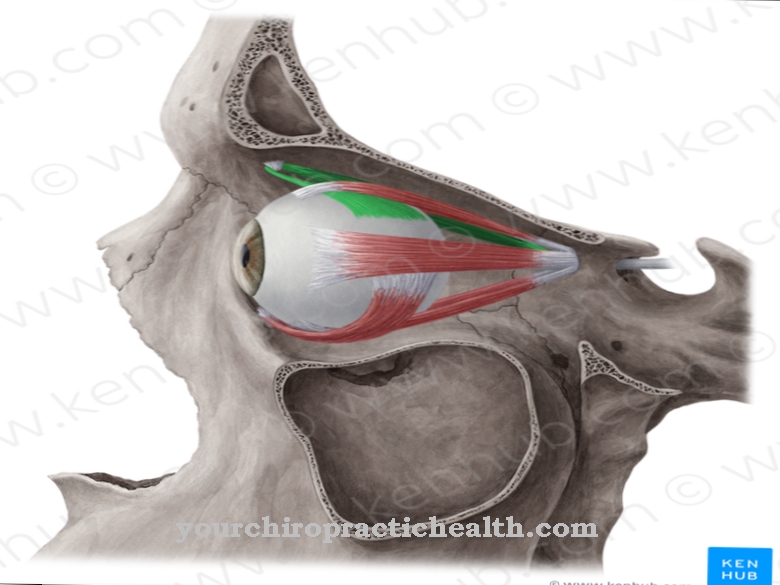
The rear of the lens rests against the front of the vitreous humor and, together with the iris, closes the anterior chamber of the eye. Around the equator of the lens capsule, zonular fibers protrude in a star shape like spokes from a wheel hub. The other end of the fibers is connected to the ciliary body, which is part of the choroid membrane of the eye as an annular bulge around the lens.
The ciliary muscle is embedded in the ciliary body and, when tensed, leads to a narrowing of the inner diameter of the ciliary body. The lens itself consists of the lens nucleus, the lens cortex and the lens capsule. The lens consists of about 60% of crystalline proteins, which are highly stable and largely UV-resistant.
A high proportion of vitamin C and oxidative stress-relieving enzymes largely prevents cloudiness from UV damage. The epithelium at the equator of the capsule produces lens fibers for life, which attach to the old fibers with loss of organelles, so that the lens enlarges and becomes less elastic in the course of life. The veinless and nerve-free lens is supplied by the aqueous humor that is formed in the ciliary body.
Function & tasks
The lens has the task of bundling incident light in such a way that a sharp image is created on the retina at the point of sharpest vision, the fovea centralis. In order to achieve a sharp image with changing distances, either the distance from the lens to the retina would have to be variable (e.g. telescope) or the focal length of the lens itself would have to be variable.
With us humans and with all vertebrates, evolution - unlike fish and reptiles - has opted for the latter variant and created the possibility of making the focal length variable within certain limits. In a mechanical second function, the lens fulfills the task of separating the anterior from the posterior eye chamber together with the iris, so that the chamber fluid cannot pass unhindered from the posterior to the anterior chamber and vice versa.
You can find your medication here
➔ Medicines for eye infectionsIllnesses & ailments
The most common lens malfunction is clouding of the lens. Another functional disorder can be caused by a mechanical displacement of the lens, a dislocation. A clouding of the lens called a cataract or cataract can have a number of causes.
The most common manifestation is the old age cataract, which only occurs at an advanced age. An inherited genetic disposition plays a role in many cases. External factors that can promote the development of cataracts are, for example, years of exposure of unprotected eyes to UV-rich sunlight at sea, in high mountains or in aircraft.
Medicines such as cortisone, drug use (including alcohol) and diabetes mellitus as well as neurodermatitis can cause the disease. If pregnant women are infected with rubella or mumps around the third month of pregnancy, there is a risk that the newborn will develop cataracts.
The disease manifests itself initially through difficulties in accommodation, later through increased sensitivity to glare and, in a more advanced stage, through clouding of vision (gray haze). The disease can be recognized from the outside by the gray color of the pupil.
A further malfunction of the lens can occur if the lens capsule is damaged in such a way that aqueous humor penetrates the lens and causes the lens cortex to swell, which leads to accommodation problems and can cause further damage in the medium term. Dislocation of the lens can result from force or from lesions of the zonular fibers.
A tumor in the ciliary body can be the cause or inherited genetic defects can lead to malfunction of the zonular fibers. A complete dislocation is when the lens either slips completely into the anterior chamber of the eye, i.e. in front of the iris, or is completely immersed in the vitreous humor. Incomplete dislocations can possibly remain symptom-free. In the case of more severe dislocations, double mono-ocular images may appear that persist when the other eye is closed or covered.



.jpg)

.jpg)
.jpg)


.jpg)