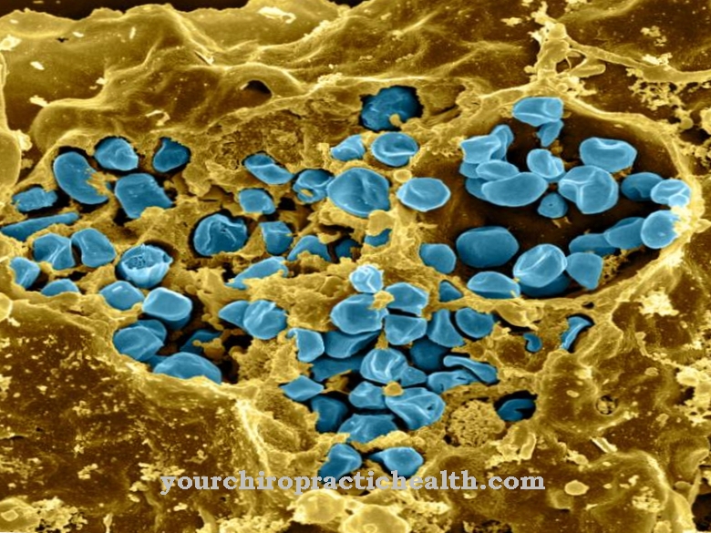
Megakaryocytes are the precursor cells of thrombocytes (blood platelets). They are located in the bone marrow and are made from pluripotent stem cells. Disturbances in the formation of platelets lead either to thrombocythemias (uncontrolled platelet formation) or thrombocytopenia (reduced platelet formation).
What are megakaryocytes?
Megakaryocytes are blood-forming cells of the bone marrow and are the precursor cells of platelets. They are among the largest cells in the human body. They can reach a diameter of up to 0.1 mm. The starting cells of the megakaryocytes are the so-called megakaryoblasts, which can no longer divide through mitosis. Instead, endomitoses are constantly taking place, which lead to the polyploid cell nuclei of the megakaryocytes. Megakaryocytes can have up to 64 times the chromosome set of normal cells. The cytoplasm of the megakaryoblasts is basophilic.
It can be colored purple or blue with basic dyes such as methylene blue, hematoxylin, toluidine blue or thionin. After several endomitoses, the mature megakaryocyte develops, the cytoplasm of which is azurophilic. The megakaryocytes represent only one percent of the blood-forming cells of the red bone marrow. A small number of megakaryocytes are also present in the circulating blood, but most of them are filtered out in the pulmonary capillaries.
Anatomy & structure
Megakaryocytes are originally formed from pluripotent stem cells. Pluripotent stem cells are embryonic cells of the bone marrow that can still differentiate into all organs of the body. From these stem cells, megakaryoblasts develop which can no longer divide through mitosis. However, constant endomitoses take place, which ultimately lead to mature megakaryocytes.
In endomitosis, only chromatids divide, but not the nuclei and cells. In this way, the cell grows more and more and forms polyploid sets of chromosomes. A 64-fold set of chromosomes can develop in the process. However, 128-fold chromosome sets were also observed. As the number of chromosomes increases, megakaryocytes become the largest cells in the bone marrow. They can reach a diameter of 35 to 150 microns. With a light microscope it looks like there are several nuclei, as the nucleus is irregularly lobed and contains coarse-grained chromatin.
The cytoplasm of the megakaryocytes is characterized by a large number of mitochondria and ribosomes as well as by a huge Golgi apparatus and a pronounced endoplasmic reticulum. In addition, the same granules are present as in the platelets. They are alpha granules, lysosomes, and electron-dense granules. These granules contain the active ingredients and proteins that stimulate platelet formation. These include growth and clotting factors, calcium, ADP and ATP.
Function & tasks
Megakaryocytes are the starting cells for the formation of platelets. The platelets are also known as platelets. When activated, they release substances to stop bleeding. After an injury, aggregation and adhesion of the platelets takes place. The injured area is sealed by the formation of fibrin and the bleeding stops. The blood platelets are small cells without a nucleus. But RNA and various cell organelles are present and enable the biosynthesis of active substances for hemostasis.
The entire process from the formation of platelets from pluripotent stem cells via megakaryoblasts and megakaryocytes is known as thrombopoiesis. First, the myeloid stem cell (hemocytoplast) develops receptors for the hormone thrombopoietin. When these receptors have formed, the hemocytoplast becomes a megakaryoblast. The hormone thrombopoietin docks on the receptor and causes endomitosis, in which only a division of the chromatin takes place, but not of the cell nucleus and the cell. The ever-growing cell develops into mature megakaryocytes, with the constant constriction of probes. Four to eight platelets can be formed per cell.
One platelet in turn produces 1,000 platelets. Therefore, between 4,000 and 8,000 platelets can develop from a megakaryocyte. The hormone thrombopoietin is absorbed through the receptors of megakaryoblasts and megakaryocytes and constantly forms platelets under endomitosis. The hormone is broken down again within the megakaryocytes and platelets.
Thrombopoietin forms in the liver, kidney and bone marrow. Since thrombopoietin breaks down within the megakaryocytes and platelets, a high thrombopoietin concentration in the blood correlates with a low megakaryocyte and platelet concentration. This stops the synthesis of the hormone. If the number of megakaryocytes and thrombocytes increases, the synthesis of thrombopoietin is stimulated again by the decrease in its concentration in the blood.
You can find your medication here
➔ Drugs for wound treatment and injuriesDiseases
Disturbances in the regulatory mechanism can lead to uncontrolled platelet formation from the megakaryocytes. This condition is known as essential thrombocytemia. In essential thrombocytemia, the concentration of platelets in the blood can reach up to 500,000 per microliter. The normal value is 150,000 to 350,000 per microliter. The cause is assumed to be the increased sensitivity of megakaryocytes to the hormone thrombopoietin.
There are abnormally large, mature megakaryocytes in the bone marrow. The clinical picture is characterized by microcirculation disorders and functional complaints. There is an increased risk of strokes and heart attacks due to thromboembolism. Insufficient blood flow to important areas of the body can lead to pain when walking, emptiness in the head or visual disturbances. In addition, upper abdominal pain caused by an enlarged liver or spleen can occur. Decreased blood platelet production is known as thrombocytopenia.
Among other things, their cause can be a disturbed formation of platelets in the bone marrow. Thrombocytopenia only becomes noticeable at a platelet concentration of 80,000 per microliter through an increased tendency to bleed. Frequent bruises, petechiae of the skin, nosebleeds or cerebral haemorrhages are to be expected.













.jpg)


