Of the Omohyoideus muscle belongs to the hyoid muscles. It also represents an auxiliary breathing muscle and is involved in chewing.
What is the omohyoideus muscle?
The lower hyoid muscles are also known as the infrahyoid muscles and include not only the omohyoideus muscle, but also the levator glandulae thyroideae muscle, the sternohyoideus muscle, the sternothyroid muscle and the thyrohyoid muscle.
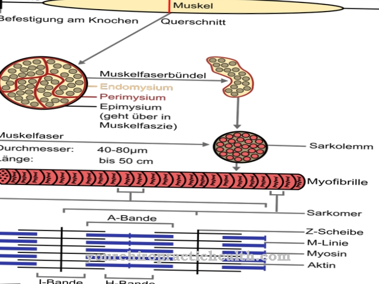
These five muscles are involved in swallowing and belong to the striated skeletal muscles. The striated pattern goes back to the structure of the tissue: Within a muscle there are numerous muscle fibers (muscle cells), each of which consists of several myofibrils. These can be divided into transverse sections, which the anatomy calls sarcomeres.
A sarcomere is bounded by the Z-disks and has two types of filaments. These are, on the one hand, strands of myosin and, on the other hand, a complex of tropomyosin and actin. These two protein structures are arranged alternately and can slide into one another, causing the muscle to shorten and thus contract.
Anatomy & structure
The omohyoideus muscle has an intermediate tendon that connects the two bellies of the muscle. It arises from the shoulder blade (scapula) and attaches to the lower hyoid bone (os hyoideum).
In anatomy, the upper part of the omohyoideus muscle is also called the superior venter ("upper abdomen"). This muscle belly is located near the sternohyoideus muscle, which, like the omohyoideus muscle, belongs to the infrahyoid muscles. The inferior venter ("upper abdomen") of the omohyoideus muscle extends upward in the neck.
In the fine structure, the omohyoideus muscle consists of muscle fibers that correspond to the muscle cells and contain many cell nuclei. The muscle fiber is surrounded by a membrane that separates it from neighboring tissue. Small tubes, the T-tubules, run through the membrane and are arranged at the level of the Z-disks of the sarcomeres. Within the membrane there are several myofibrils, which are thread-like strands. Analogous to the endoplasmic reticulum in other cell types, the sarcoplasmic reticulum is located in the interstices. Mitochondria are responsible for cell respiration and play a central role in energy metabolism, which is why they are also known as the “power plants of cells”.
Function & tasks
The ansa cervicalis profunda connects the omohyoideus muscle with the nervous system and controls its activity. The ansa cervicalis profunda is the deep part of the cervical nerve loop, which also innervates the other muscles of the infrahyoid muscles. The signals from the nerve loop come from the cervical nerve plexus (cervical plexus).The ansa cervicalis profunda runs across the internal jugular vein.
Oxygen-depleted blood flows through this vein from the head back towards the lungs. The omohyoideus muscle is responsible for keeping the internal jugular vein open by tensing the middle cervical fascia (lamina praetrachealis).
The omohyoideus muscle can also pull the hyoid bone down and back by contracting it. This movement is particularly relevant when swallowing. Various muscles work together in this process: In addition to the infrahyoid muscles, the muscles of the floor of the mouth (suprahyoid muscles) and the muscles of the palate are also active when swallowing. Then the tunica muscularis of the esophagus supports the transport of food or fluid into the stomach. The swallowing center in the elongated medulla (medulla oblongata) coordinates the swallowing process and triggers the swallowing reflex.
The swallowing center receives sensitive information via the ninth and tenth cranial nerves (glossopharyngeal nerves and vagus nerves) and uses various nerve pathways to trigger the motor reaction in the corresponding muscles. The neural structures involved include the ninth to twelfth cranial nerves, the fifth cranial nerve and the cervical plexus, which also controls the omohyoideus muscle.
Furthermore, the omohyoideus muscle participates in certain head movements. When a person moves their head forward, the infrahyoid muscles (including the omohyoideus muscle) cause the hyoid bone to move backwards and downwards. In its function as an auxiliary breathing muscle, the omohyoideus muscle also supports breathing to a lesser extent.
You can find your medication here
➔ Medication for shortness of breath and lung problemsDiseases
One of the tasks of the omohyoideus muscle is to keep the internal jugular vein open. Doctors sometimes use this vein to create a central venous catheter (CVC). To do this, they insert a thin tube into the vein and push it inside the blood vessel up to the right atrium.
A central venous catheter is used in various situations. For example, doctors can use it to determine the central venous pressure, which is an indicator of the preload of the heart and plays an important role in various cardiological diseases. In addition, a CVC allows various substances to be administered close to the heart, including electrolytes and medication. For the central venous catheter, not only the internal jugular vein comes into consideration, but also the subclavian vein. In addition to these two preferred CVC variants, access can also be made into the anonymous vein or the basilic vein, and more rarely via other veins.
Damage and functional limitations of the omohyoid muscle can contribute to swallowing disorders. Neurological disorders, for example in connection with a stroke or a neurodegenerative disease, can damage the nerve fibers that are responsible for supplying the omohyoideus muscle and the other infrahyoid muscles. Injuries, tumors and other lesions in the elongated medulla (medulla oblongata) can also affect the swallowing center and hinder the coordinated swallowing process. The swallowing reflex may also be affected.



.jpg)

.jpg)
.jpg)


.jpg)