The Orbit is the bony eye socket. Seven bones meet in this receptacle for the eye. The weakest point of the orbit is the floor, which is often affected by fractures after being hit.
What is the orbit
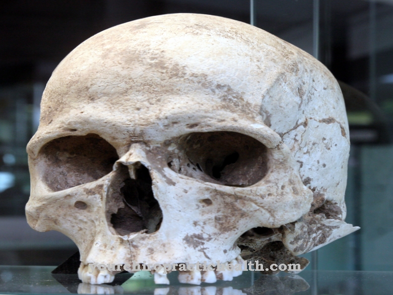
The orbits are the bony eye sockets. These are pits four to five centimeters deep in the skull in which the eyes and their attachments are located. These pits each consist of seven bones. In addition to the frontal bone, the lacrimal bone and the upper jaw, the cheekbone, ethmoid and palatine bone meet here. In addition to the bony eye socket, the tear bone is also involved in the nasal bone.
The frontal bone is the anterior roof of the skull and thus the upper wall of the skull cavity. The upper jaw borders both the oral cavities and the nasal and eye sockets. The cheekbone is a pair of facial bones and the ethmoid bone delimits the cranial cavity at the end of the nasal cavity from the face. The palatine bone is primarily involved in the nasal and oral cavities. The sphenoid bone is again a cranial skull bone in the lower central area, where it forms the rear part of the orbit. Inside the orbit are several holes through which nerves and blood vessels of the eyes and face pass. Around 4/5 of the orbits consist of fat, connective tissue, muscles, nerves and vessels. The last fifth is the eyeball.
Anatomy & structure
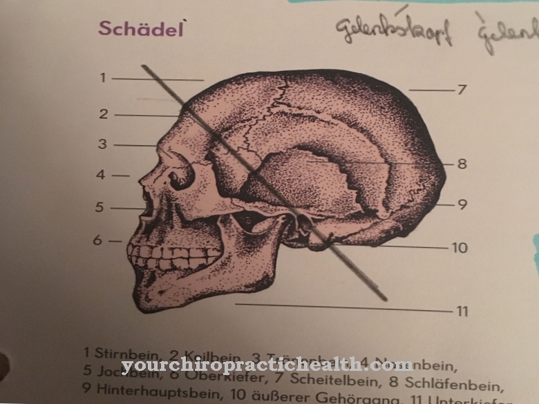
The frontal and sphenoid bones form the roof of each eye socket. The maxilla, the os zygomaticum, and the os palatinum each form the orbital floor. The lateral wall is formed by the os zygomaticum and the os sphenoidale, while the maxilla, the os lacrimale, the os ethmoidale and the facies orbitalis ossis frontalis together with the ala minor ossis sphenoidalis form the middle wall of the orbit. The structure of meeting bones in each orbit has the shape of a four-sided pyramid. The base of this pyramid faces forward. The point points into the depths of the skull.
The contents of the orbit are separated from the bones by the periorbital layer of tissue. Frontally, the bony eye sockets have an access called the aditus orbitalis, which is bordered by the bony orbital rim. There is a connection between the orbit and the middle fossa with the fissura orbitalis superior and the canalis opticus. This is where conduction pathways enter the eye sockets. Many nerves and vessels also pass through the infraorbital sulcus, which forms an entrance to the infraorbital canal. Nerves and blood vessels recede into the cranial cavity through the foramen ethmoidale anterius and the foramen ethmoidale posterius.
Function & tasks
The orbits are the receptacles for the eyes and their supply lines made of blood vessels and nerves. They also serve the bony protection of the eye. Since the eye socket is around five centimeters deep, the eyeball and its supply structures are not damaged as easily as if they were lying flat on the face. The seven adjoining bones of the orbit enclose and even completely protect the eyeball on three sides.
In addition to the bones, the periorbitae, the fat and the connective tissue of the eye sockets play a particularly protective role. The holes in the orbit mean that nerves such as the optic nerve can pass through. In this respect, the bony eye sockets also take on the tasks of a supply structure guide rail. In addition to the optic nerve, the ophthalmic artery, the inferior ophthalmic vein, the lacrimal ducts, the zygomatic nerve and the infraorbital nerve are led from here.
The orbital fissure also leads to the cranial nerves of the eye muscles and the sensitive bulb. These cranial nerves include the third cranial nervus oculomotorius, the fourth cranial nervus trochlearis and the first fifth cranial nerve ophthalmic nerve and the sixth cranial nerve abducens nerve. The eye socket also offers these structures protection and additional stability. Some structures of the bony eye socket are stronger than others and thus guarantee better protection. The weaker structures include the inner side wall and the bottom of the eye sockets. These weaker parts play a role especially in connection with fractures.
You can find your medication here
➔ Medicines for eye infectionsDiseases
Problems with the orbits are usually the result of a blow to the eye. Often in such scenarios, the weak parts of the orbit are affected by fractures. One of the most common phenomena is the orbital floor rupture, in which the eye socket breaks through to the maxillary sinus. A break in the orbital floor is usually expressed in double images, which can be traced back to a restricted movement of the eye.
Muscle tissue is often pinched in the hernia. Connective and holding tissue, and more rarely nerve tissue, slip into it just as often. As soon as nerve tissue is affected, sensory disorders in the face can add to the double vision. Fractures in the orbital floor can be treated surgically. Such reconstructive treatments of the eye socket take place especially when muscles or nerves are trapped, as the jammed structures could otherwise die off. Especially the liberation of nerves from a fracture gap can still permanently damage the jammed nerve.
As part of the reconstructive operation, the patient is usually given a tiny metal plate that holds the floor of the eye socket together and thus helps it grow together. The plate can, but does not have to be, removed again. With an untreated orbital floor fracture, the eye can sag a little in the worst case. Sometimes the orbits are also affected by inflammation or cysts. However, fractures remain the most common symptoms.




.jpg)

.jpg)
.jpg)


.jpg)