The Tunica media is part of the walls of blood and lymph vessels, which lies between two other layers. It contains, among other things, muscle cells with which the body regulates the width of the veins. Damage to the tunica media can narrow the blood vessels (arteriosclerosis).
What is the tunica media?
The tunica media is part of the vein and artery walls. In order to differentiate it from the middle skin of the eye (tunica media bulbi or uvea), doctors sometimes refer to the middle vascular layer as the tunica media vasorum.
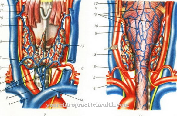
It is surrounded by the tunica adventitia or tunica externa. The tunica externa forms the outermost wall of the blood vessels. The tunica lies intima on the inside of the arteries and veins. The wall of the lymph vessels also has a tunica media in the middle. The tissue of the tunica media is not made up of a uniform structure, but is made up of muscle cells, collagen, elastic fibers and connective tissue. The muscle cells are particularly important for the transport of fluid in the vessels. With age, the elasticity of the vascular walls decreases and can lead to normative constrictions.
Anatomy & structure
Some cells in the vessel wall are muscle cells. Since the larger arteries have to pump the blood through the organism, they have a thicker tunica media. The extra muscle cells help the blood vessels build up the necessary pressure.
In between there is collagen, a special protein molecule, and elastic fibers. The latter give the fabric its flexibility. In addition, the tunica media consists of connective tissue that supports the other cells and keeps them in shape. The connective tissue also takes on a supplying role: it transfers nutrients and oxygen to the other cells and distributes the resources.
Medical professionals classify arteries into different types; the differences are also reflected in the tunica media. For example, the muscular arteries have stronger muscles, while the elastic arteries have more elastic fibers and collagen.
Function & tasks
The tunica media makes a decisive contribution to ensuring that the blood flows evenly through the human body. In the arteries, blood flows away from the heart. In the lungs, the red blood cells take up and distribute oxygen. The heart serves as a pump. But the arteries themselves also have to drive the blood to keep it flowing.
In the larger arteries, people can easily feel the rhythmic pumping; the blood vessels are therefore also called arteries. When the arteries are injured, the blood often shoots out of the wound, which shows the high pressure inside the vessel. In order for the arteries to perform their pumping movements, they need muscles. The muscle layer is located in the tunica media and forms a ring around the arteries. The muscle cells in the tunica media belong to the smooth muscles and thus belong to the same fiber type as the heart muscle. People cannot consciously control or suppress these movements.
Not only blood vessels have a vessel wall with a tunica media; lymph vessels are also dependent on them. The lymph vessels collect fluid from the spaces between the cells. They appear in almost every major tissue. Similar to blood vessels, they can be of different sizes and flow into one another. Eventually, the lymph vessels release the collected fluid into the blood vessels.
The body excretes excess fluid in the urine. The lymphatic system is used to transport fluids and ensures that no water collects between the cells. The lymphatic vessels also carry certain macromolecules - for example proteins and lymphocytes that are part of the immune system.
You can find your medication here
➔ Medicines against swelling of the lymph nodesDiseases
The tunica media can, among other things, be involved in the development of arteriosclerosis. This is a blockage of the bloodstream, for which various causes can be responsible. For example, blood lipids, called triglycerides, can form clumps in the arteries and veins as the molecules deposit on the vessel walls. This leaves less room for the bloodstream to pass through the affected area.
The risk of such deposits is particularly high on vascular valves and in finer veins. As a result of a vascular blockage, the body can no longer supply the underlying tissue with oxygen and nutrients. The removal of carbon dioxide, other waste materials and cell products is also disturbed by arteriosclerosis. In addition, the deposits can tear themselves away and get into other parts of the body with the bloodstream.
They either dissolve or close the vessels in which they get stuck. In this way, blockage of the arteries potentially leads to stroke, heart attack, or pulmonary embolism; Other tissue can also be affected by the arteriosclerosis and, in the worst case, die.
Proper muscle movement of the arteries is also required to keep the arteries from occluding. The tunica media contains smooth muscles that allow the blood vessels to widen or narrow as needed. High blood pressure (hypertension) can damage the tunica media: the cells in the vessel wall receive little oxygen and die: the regulation of the arterial width is disturbed and the artery can narrow to the point that arteriosclerosis is present.
In Mönckeberg's sclerosis, calcium is deposited in the tunica media and also leads to functional restrictions in the blood vessels.

.jpg)









.jpg)
