Of the Lower jaw (lat. Mandible) is part of the human facial skull. Together with the upper jaw, it forms the chewing apparatus. The upper jaw represents the immobile part and the lower jaw the movable part during the chewing process.
What is the lower jaw?
The lower jaw of humans will too Jaw called. It is one of the bones of the facial skull. Its Latin name mandibula is derived from the Latin word “mandere” for “chew”. This is due to its crucial role in the chewing process. In contrast to the upper jaw, which is firmly fused with the other cranial bones, the human lower jaw is mobile.
It is connected to the upper jaw by the temporomandibular joint. Therefore it can be opened and closed by the masticatory muscles and also moved sideways in both directions. During embryonic development, the lower jaw arises from the first gill arch. The mandibular nerve supplying it develops analogously from the first branchial arch nerve.
Anatomy & structure
The actual mandibular body, Corpus mandibulae, resembles a horseshoe in its curved shape. The center of the arch supports the chin. The lower jaw has an ascending branch of the lower jaw (Ramus mandibulae) on both sides. On each branch of the lower jaw there is a muscle extension (coronoid process), which leads to the insertion of the temporal muscle. The branches of the lower jaw open into the articular processus condylaris.
There is a notch (Incisura mandibulae) between the muscle process and the articular process. The joint process together with the temporomandibular head of the temporomandibular joint creates the temporomandibular articulation. Between the temporomandibular joint head and the joint socket on the skull there is a movable, buffering cartilage disc (discus). The jaw muscles also attach to the branches of the lower jaw. There are four paired muscles: the masseter muscle, the temporal muscle, the medial pterygoideus muscle, and the lateral pterygoideus muscle, the internal wing muscle.
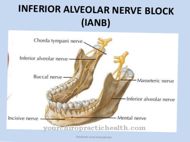
There is a bone tongue (lingula mandibulae) on the inside of each branch of the lower jaw. It covers the mandibular foramen. The nerve of the mandibular tooth fan (nervus alveolaris inferior) enters the mandibular canal (canalis mandibulae) through the mandibular holes. This nerve is an extension of the mandibular nerve of the mandibular nerve The inferior alveolar nerve runs under the tips of the roots of the posterior teeth. Its terminal branch is the chin nerve, the mental nerve. It emerges from the lower jaw body through the mental foramen in the area of the root tips of the premolars.
Other nerves located in the lower jaw are the masseteric nerve, the deep temporal nerve, the medial pterygoid nerve and the lateral pterygoid nerve. The lower jaw hole also acts as a passage point for the lower dental artery inferior alveolar artery and the associated inferior alveolar vein.
Function & tasks
In general, the lower jaw has the task of closing off the oral cavity and performing the chewing movements. This is only possible because the TMJ allows it to move in all directions. In addition, it is also necessary for the generation of specific sounds, such as human speech.
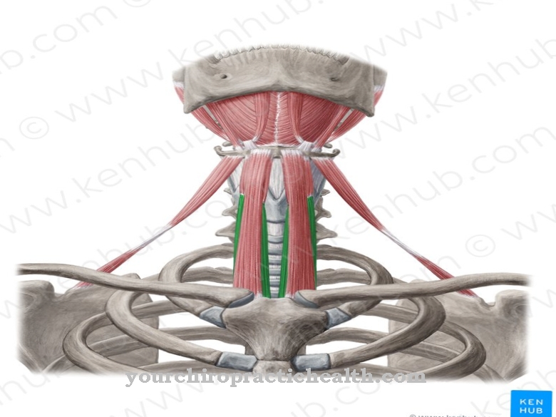
The four pairs of muscles in the lower jaw each have specific, complementary functions. The jaw muscle (M. masseter) serves to close the jaw, as does the temple muscle (M. temporalis). The latter is also needed to retract the lower jaw. The inner wing muscle (M. pterygoideus medialis) also helps to close the jaw. The outer wing muscle (M. pterygoideus lateralis) causes the opening and advancement of the lower jaw. In addition, it implements the grinding slide movements to the left and right.
The nerves of the lower jaw are also assigned to precise functional areas: The inferior alveolar nerve renervates the teeth and tooth sockets of the lower jaw. The chin nerve supplies the skin of the chin and lower lip. The masticatory nerve (N. massetericus) transmits information to and from the masseter muscle. The temporal nerves supply the temporal muscles. The inner and outer wing muscles are each innervated by the nervus pterygoideus medialis and lateralis. The lower dental artery (A. alveolaris inferior) and the respective vein (V. alveolaris inferior) serve to supply the lower jaw with blood.
You can find your medication here
➔ Toothache medicationDiseases
Complaints of the lower jaw almost always occur in combination with disorders of the entire chewing apparatus. The finding of complaints in the chewing apparatus in Germany is therefore often craniomandibular dysfunction (CMD). This is a summary of disorders from the functional, structural, psychological and biochemical areas. It manifests itself in dysregulation of the joint and muscle functions of the temporomandibular joints.
Due to its broad and often combined causes, CMD manifests itself in numerous symptoms: The jaw joints can rub or crack when opening and closing. Sometimes the ability to open the jaw wide is severely restricted. This can also lead to problems with biting off, laughing and speaking. Pain emanating from the jaw can radiate to the teeth, the entire oral cavity, face, head, neck and shoulder area and the spine.
Sometimes the teeth suddenly don't fit properly. Earache, tinnitus and dizziness can also be caused by (lower) jaw problems. Likewise, eye problems and difficulty swallowing. In general, the (lower) jaw complaints are assumed to have either ascending or descending symptoms. With the ascending symptoms, displacements of the spine affect the cervical spine and from there on the temporomandibular joints. With the descending symptoms, imbalances in the jaw area lead to complaints in the neck, shoulders and spine.
The causes of disorders in the (lower) jaw area are diverse. They can lie in bacterial and viral inflammation of the jawbones and temporomandibular joints. A common cause is also nocturnal teeth grinding (bruxism) due to mental tension and / or misaligned teeth. Since the temporomandibular joint is the most frequently used joint in the body, signs of wear and tear can occur with age (TMJ osteoarthritis). A shift in the cartilage disc of the temporomandibular joint can also cause pain and signs of wear and tear.
The dentist is initially the contact for all disorders summarized in the CMD. If necessary, he will then request further, for example (orthodontic) orthopedic services.










.jpg)
