The Azygos vein begins above the diaphragm and is a branch of the lumbar vein (ascending lumbar vein). It transports oxygen-poor blood to the heart. In the event of a drainage disorder, the azygous vein can contribute to a bypass circuit because of its connections to other veins.
What is the azygous vein?
The azygos vein is an ascending vein in the trunk of the human body that originates from the lumbar vein (ascending lumbar vein) and merges into the superior vena cava (superior vena cava).
The azygous vein is part of the body's circulatory system, also known as the great bloodstream, and it carries oxygen-poor blood. The term “vein” is derived from the Latin verb “venire”, which means “to come”. The blood flow from the veins leads to the heart, whereas arteries carry the blood away from the heart. These names are independent of whether the veins transport blood that is poor in oxygen (as in the body's circulation) or oxygen-rich (as in the pulmonary circulation).
The azygos vein represents an unpaired vein, as it has no exact equivalent in the left half of the body. The hemiazygos vein on the left follows a different course than the azygos vein on the right. The azygos vein owes its name to this fact, which goes back to the Greek word for “unpaired”.
Anatomy & structure
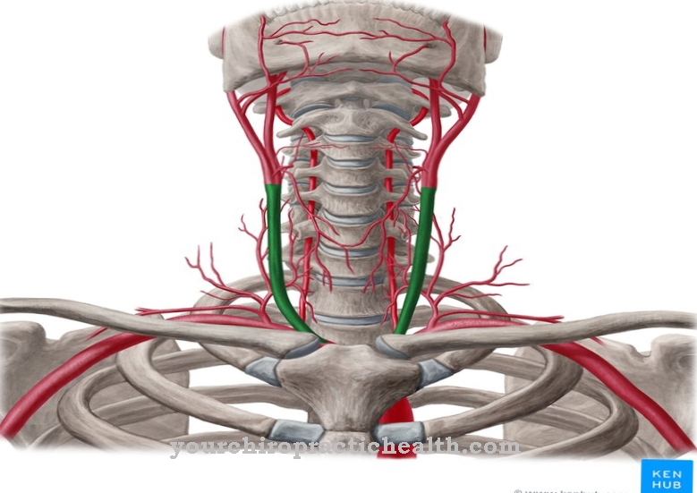
The origin of the azygos vein is in the right lumbar vein (ascending lumbar vein). The azygous vein branches off from this blood vessel above the diaphragm. The azygos vein runs on the right side of the spine, while the hemiazygos vein extends on the left.
The bronchial veins (venae bronchiales) and the intercostal veins (venae intercostales posteriores) open into the vena azygos. In addition, the blood flows from the esophageal veins (Venae oesophageales) and from the Vena hemiazygos into the Vena azygos, which in turn ends in the superior vena cava (Vena cava superior). Before the vena azygos merges into the superior vena cava, it runs in an arc, which medicine also calls the arcus venae azygos.
The wall of the azygos vein consists of three layers, with the intima being the innermost of them. The tunica media lies in the middle, but is not clearly delimited from the outer tunica externa. In general, the walls of veins are thinner than the walls of arteries and, above all, they have less pronounced (smooth) circular muscles in the tunica media.
Function & tasks
The task of the azygous vein is to take in oxygen-poor blood from various confluent vessels and to transport it to the superior vena cava. From there the blood continues to flow into the right atrium. The vital organ then pumps the blood into the right ventricle, bringing it to the pulmonary circulation, also known as the small bloodstream. Via the pulmonary trunk (Truncus pulmonalis), the oxygen-poor blood finally reaches the lungs, where oxygen can bind to the red blood cells (erythrocytes).
The blood of the azygos vein comes from the bronchial veins, among other things, which divert blood from the bronchi and bronchial lymph nodes. Lymph nodes belong to the lymphatic organs and as such embody a part of the immune system, which is dedicated to fighting diseases and pathogens. The venae intercostales posteriores also form tributaries to the vena azygos. These blood vessels form a group of intercostal veins, they are the counterpart to the intercostal arteries. The posterior intercostal veins represent the posterior intercostal veins and drain blood from the intercostal space, which anatomy also calls the intercostal space or space intercostale.
Not all intercostal veins flow into the azygos vein; instead, some of them also open into the hemiazygos veins and the internal thoracic veins. The oesophageal veins surround the esophagus and supply the azygos vein with blood that has previously been enriched with oxygen via the aorta, intercostal artery, thyroid artery and left gastric artery.
You can find your medication here
➔ Medication for heartburn and bloatingDiseases
Drainage disorders affect the blood flow in venous vessels. If such a drainage disorder affects the superior or inferior vena cava (superior vena cava or inferior vena cava), the azygos vein can contribute to the bypass circuit - comparable to the function of an artificial bypass.
Medicine calls connections between blood vessels anastomoses. Various causes come into question for the development of a drainage disorder, which also determine possible treatment options. Tumors that narrow the veins are a potential cause. Thrombi and other debris within the vein can also cause it to narrow. Vein weakness or venous insufficiency particularly often affects the lower extremities and often manifests itself in symptoms such as swollen legs or feet, visible veins and spider veins, skin changes and pain in the legs. Symptoms may vary if there is an obstruction in the upper veins.
A dilation of the azygos vein is also one of the indicators (of several) for pulmonary hypertension. This is increased blood pressure in the pulmonary circulation, which is based on increased vascular resistance. The expansion of the azygos vein is one of the X-ray signs of the clinical picture: A vein diameter of over 7 mm is considered critical. Pulmonary hypertension can be based on various chronic and acute courses, for which, for example, structural changes or physiological stress reactions can be responsible. In this case, too, treatment depends on the cause.

