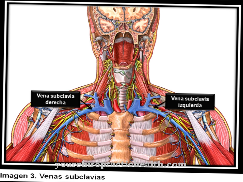
The Subclavian vein, also Clavicle vein called, runs behind the collarbone above the first rib. It transports the blood in the arm towards the heart.
What is the subclavian vein?
The subclavian vein belongs to the veins of the small body circulation in the area of the arm and neck. A distinction is made between the right and left subclavian veins. It is one of the root veins of the brachiocephalic vein.
Primarily, it transports blood from the upper extremities with arm and shoulder and reaches the right atrium via the vein angle through the vena brachiocephalica (head and arm vein). From there, the blood flows through the pulmonary circulation (small circulation) into the lungs and is enriched with oxygen. The oxygen-enriched blood flows into the left atrium and from there is pumped back into the body through the aorta (large body artery) to supply the tissue with oxygen (large blood circulation).
Almost all arteries carry oxygen-rich blood and most veins carry oxygen-depleted blood. The venous blood is dark red compared to the arterial blood because the oxygen has been withdrawn. The blood pressure in the veins is significantly lower than that in the arteries and is called the low pressure system of the blood circulation.
Anatomy & structure
The subclavian vein is only a few centimeters long and runs horizontally towards the middle of the body. It is an accompanying vein that runs parallel to its corresponding artery (arteria subclavian-subclavian artery).
Another paired artery, which carries the oxygenated blood from the heart back to the head, neck, arm and shoulder. The subclavian vein is the direct continuation of the axillary vein (axillary vein). This in turn is a continuation of the brachial vein (arm vein), whereby the transition is not clearly defined anatomically. Together, the subclavian vein and the axillary vein form the main trunk of the arm veins. The subclavian vein and the internal jugular vein (internal jugular vein, vein of the neck), which are important for the blood outflow of the brain, are both root veins.
They unite in the vein angle to form the brachiocephalic vein (head-arm vein). It is also a paired body vein, the slightly shorter right part meeting the left brachiocephalic vein at the level of the first costal cartilage. Here both veins join to form the superior vena cava (superior vena cava), which ends in the right atrium. It is the largest vein in the human body.The subclavian vein is firmly connected to a covering layer of connective tissue (fascia clavipectoralis) on the periosteum of the clavicle. This prevents the vein from collapsing (collapse) and facilitates the suction of blood from the outer zone of the body (periphery) when the arm and shoulder are moved.
Function & tasks
The subclavian vein is responsible for transporting the oxygen-poor blood of the arms, shoulders and the lateral chest wall. The blood flow runs over the vein angle, to the head and arm vein and finally through the superior vena cava to the right ventricle. From there, the blood is pumped through the pulmonary valves into the pulmonary artery and then into the lungs.
In the lungs, the blood is enriched with oxygen and flows back through the mitral valve into the left ventricle. From there via the aortic valve into the main artery (aorta) in order to finally distribute the oxygen-rich blood via the capillaries in the body. The subclavian vein receives its inflows from the external jugular vein (external jugular vein), which forms behind the ear through the union of the occipital vein (occipital vein) and the auricular vein (ear vein). It receives further inflows through the accompanying veins of the subclavian artery.
There are functional differences between the right and left subclavian veins. The left side is a little more important, because here, among other things, the lymphatic trunk flows in, which transports lymph from the entire lower half of the body. Ani on the right side is a small lymph vessel that carries lymph from the right arm, the right side of the chest, and the right side of the neck. The lymphatic system specializes in the transport of nutrients and waste materials and, along with the bloodstream, forms the most important transport system in the body.
Diseases
Thoracic outlet syndrome is a compression (squeezing) of the vascular nerve bundle, consisting of the brachial plexus (arm plexus), the subclavian artery and the subclavian vein.
This vascular nerve bundle has to overcome three narrow points on the way towards the upper extremity: the scalenus gap (denotes the gap between the rib support muscles), the costoclavicular space (space between the first rib and the clavicle) and the coracopectoral space (the space between the bone process of the shoulder blade and the small pectoral muscle). The thoracic inlet syndrome is a special form of the thoracic outlet system. It describes the narrowing of the subclavian vein and can lead to subclavian thrombosis or acute axillary vein congestion (Paget-von-Schroetter syndrome).
A subclavian vein thrombosis is rare compared to leg and pelvic thrombosis. A thrombosis is a blood clot (thrombus) that narrows or blocks the blood vessels. It occurs when not enough venous blood flows to the heart. Often the thrombosis of the subclavian vein occurs as a result of physical overexertion during sport or “overhead” activity. However, it can also occur due to a tumor or a central venous catheter.
It mostly affects young adult men. The thrombosis occurs predominantly on the right side. Phlegmasia coerulea dolens is a rather rare clinical picture. A complete occlusion of all veins in an extremity (thrombosis). The reason is a disruption of the microcirculation (part of the bloodstream of the smallest blood vessels). Phlegmasia coerules dolens is an emergency and requires rapid surgical intervention.



.jpg)

.jpg)
.jpg)


.jpg)