The Toe bones belong to the delicate structures of the human skeleton. As a freely movable part of the bony foot, they can be assigned to the lower extremity. With the exception of the two-part big toe, the toes each consist of three individual bone members.
What are the toe bones?
The toes are located at the distal end of the foot and, as its end links, form the final end of the lower extremity. Analogous to the hand, a distinction can be made between the foot, the tarsal bones, the metatarsal bones and the toe bones in the structure of the foot.
In total, the foot skeleton is composed of 26 individual bones, including 14 toe bones. These connect distally to the metatarsal bone. In all five toes, they are made up of several individual bones, the so-called phalanges. These are referred to as proximal, medial or distal phalanges or also toe basal limbs, toe limbs and toe limbs according to the position facing away from and facing the trunk of the body. The phalanges are held together by articulated connections as well as muscles, ligaments and tendons and are therefore flexibly movable.
Anatomy & structure
The individual toes of the big toe have two, otherwise all other toes have three bony limbs, i.e. phalanges. The big toe lacks the medial phalanx.
The structure of the phalanges can be divided into a proximal base, a central body and a distal head. The toe bones belong to the elongated tubular bones with two cartilaginous joint ends and an intermediate shaft. The basal phalanges of the toes, which connect directly to the tarsal bone, are longer than the middle and end phalanges and have a biconcave shape. The stocky-looking medial phalanges form the middle link between the proximal and distal phalanges. In terms of size, the middle toe phalanx is also in the middle, with the shaft slightly wider than that of the basic phalanges.
The relatively stunted and flattened end links are comparatively the shortest of the toe bones. Different foot shapes are distinguished based on the length of the toe. The most common is the so-called Egyptian foot, where the big toe is the longest.
Function & tasks
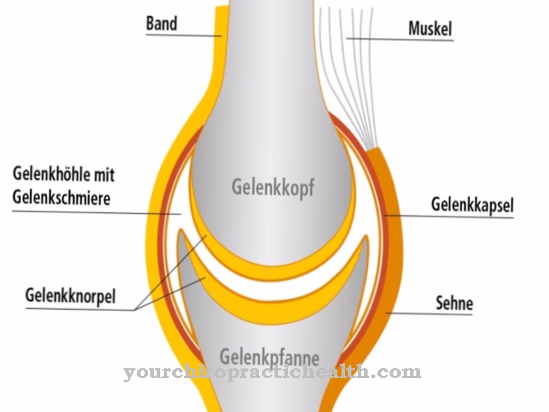
The connection of the individual phalanges is based on small joints. The articular surfaces of the metatarsophalangeal joints, also called metatarsophalangeal joints, are formed from the bones of the metatarsus and the associated metatarsals. The joints that lie between the proximal and middle phalanx as well as between the middle and terminal phalanx are described as the toe and toe joints or as proximal and distal interphalangeal joints.
All toes except for the big toe are therefore each equipped with three joints: the base joint and the two interphalangeal joints. The metatarsophalangeal joints are functionally assigned to the egg joints, which enable movement in two axes, namely ab- and adduction, i.e. Movements to the side, as well as flexion and extension, i.e. Forward and backward movements. The interphalangeal joints are hinge joints that only allow one direction of movement with flexion and extension. Due to the lack of a medial phalanx, the big toe has only one interphalangeal joint. In summary, the following movements can be carried out with the toes: bending in the direction of the sole of the foot, stretching in the direction of the back of the foot and spreading and contracting the toes.
The foot with its anatomically differentiated, freely movable toe members is the part of the human musculoskeletal system on which the various fine motor processes of locomotion are based. The stabilizing function of the toes is not only a prerequisite for running or walking, but also for certain types of sport or movements such as hopping or dancing. The big toe is essential for all of the fine motor functions. This serves both to roll and cushion the foot and to push it off the ground, i.e. to accelerate the walking movement.
Diseases
Malformed toe bones or toes that can only be stressed to a limited extent due to the disease have a limiting effect on the mobility of those affected. Different clinical pictures such as osteoarthritis and gout, but also malformations or fractures come into question as the cause of this impairment.
The clinical picture of osteoarthritis describes degenerative signs of wear and tear on joints, which can usually be traced back to a progressive destruction of the protective cartilage layer on the joint surfaces. Swelling and limited mobility in the area of the joint are symptomatic, as well as load-dependent pain at the beginning and pain at rest in the further course. As the disease progresses, incorrect posture can be observed, which causes contractures and ultimately a stiffening of the joint. The stiffening of the metatarsophalangeal joint caused by wear and tear as a consequence of osteoarthritis is known as hallux rigidus. The deformity that is most frequently noted in the area of the foot is hallux valgus or bunion.
The metatarsophalangeal joint of the big toe is bent laterally outwards and the first metatarsal shows an inward deviation. Outwardly, this is represented by a protruding ball of the toe. Deformities in the area of the little toes, i.e. toes 2-4, include the hammer toe and the claw toe. The hammer toe is characterized by a hammer-like bent toe with simultaneous hyperextension in the metatarsophalangeal joint. Depending on whether the deformity can be reduced or not, a distinction is made between a flexible and a fixed hammer toe. Typical of a clawed toe is a claw-like bent toe with simultaneous subluxation or dorsal dislocation of the metatarsophalangeal joint.
The tip of the toe usually no longer touches the ground. A fracture of the toes usually affects the distal phalanx. Most often, such a break is caused by an immediate, external force on the bone.

.jpg)









.jpg)
