The Alpha-1 fetoprotein (AFP) is mainly formed in the embryonic tissue and serves as a transport protein there. Very little AFP is produced after birth. Elevated serum or blood values in children and adults indicate, among other things, tumors.
What is Alpha-1 Fetoprotein?
Alpha-1 fetoprotein is a protein that is formed in the endodermal tissue during embryogenesis. The endodermal tissue develops from the yolk sac and forms the starting point for the development of various tissues and organs such as the digestive tract, liver, pancreas, thymus, thyroid, respiratory organs, urinary bladder or urethra.
From the fourth week of pregnancy onwards, alpha-1-fetoprotein is mainly produced in the yolk sac and in smaller quantities also by the growing liver of the fetus. Its concentration reaches its highest values in the twelfth to sixteenth week of pregnancy. Shortly after birth, the synthesis of AFP comes to an almost complete standstill. In adults and children, higher concentrations are a sign of pathological processes in the body. The alpha-1 fetoprotein, for example, serves as a tumor marker.
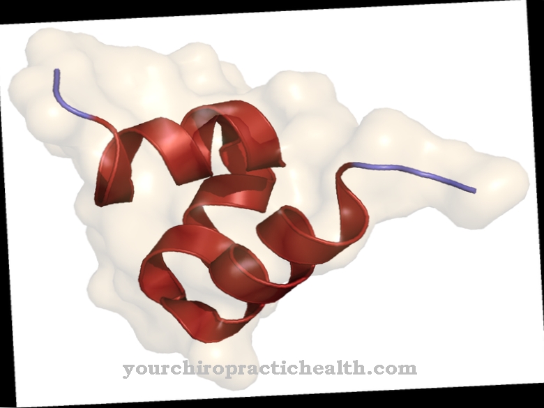
The measurement of blood and serum concentrations in pregnant women is used to diagnose neural tube defects in the fetus or to reveal Down's syndrome. The protein consists of 591 amino acids. Usually there is only one chain. Dimer or trimer protein chains are rarely found in alpha-1 fetoprotein.
Function, effect & tasks
The alpha-1 fetoprotein is of great importance for the growing embryo. This is why it is also formed in larger concentrations in embryonic tissue (especially in the yolk sac). It serves as a transport protein during embryogenesis.
It enables the trace elements nickel and copper to be transported in the fetal blood. It is also responsible for transporting bilirubin and fatty acids in the fetus's blood. Therefore, increased values in the serum, blood plasma or amniotic fluid can also be measured in pregnant women. The yolk sac of the embryo is the actual metabolic organ until the liver has formed. It needs alpha-1 fetoprotein to make the developing embryo more and more independent of the maternal blood circulation.
After birth, this protein is no longer necessary and is only synthesized in very small quantities in the digestive tract. However, alpha-1 fetoprotein production increases as tumors grow.
Education, occurrence, properties & optimal values
In non-pregnant women, men and children, the normal concentration of alpha-1 fetoprotein in the blood plasma and serum is less than seven nanograms per milliliter. However, there is a gray area up to 20 nanograms per milliliter. However, it should be mentioned that no clear limit values have been set in Germany. However, if the concentration of AFP exceeds 40 nanograms per liter, the possibility of cancer growth should also be considered.
In pregnant women, the concentrations of AFP in the blood plasma, serum and of course in the amniotic fluid are increased. The serum AFP concentration in pregnant women is always determined as part of prenatal screening tests. Here the concentrations are given as so-called MoM values. MoM means "multiple of the central value". During pregnancy, the AFP concentrations rise exceptionally and change constantly depending on the stage of pregnancy.
The AFP concentration in the serum should not exceed a value of 2.5 MoM, because elevated values may indicate a neural tube defect in the fetus. The normal value for pregnant women is 05 to 2.0 MoM. Lower levels of alpha-1 fetoprotein can in turn indicate trisomies such as Down syndrome.
Diseases & Disorders
Deviating values of alpha-1-fetoprotein in blood plasma or serum indicate pathological processes in both pregnant women and non-pregnant women as well as children and men. If the values are elevated in pregnant women, it may be a neural tube defect in the growing child.
The neural tube defect is characterized by incomplete closure of the neural tube. Large amounts of alpha-1 fetoprotein enter the pregnant woman's blood plasma or amniotic fluid through the open neural tube. If the concentration is above 2.5 MoM, these malformations should be considered and further ultrasound examinations performed. Congenital neural tube defects such as anencephaly (missing brain) or spina bifida (open back) as well as abdominal wall defects can be detected. If the concentration of AFP is below 0.5 MoM, trisomy 21 (Down syndrome) or other trisomies may also be present.
However, deviating AFP values in pregnant women only provide an indication of possible defects. Imaging methods such as ultrasound examinations in particular must confirm the diagnosis. Elevated values also occur in multiple pregnancies or in an incorrectly dated pregnancy. Targeted ultrasound examinations can be carried out in the borderline range of 2.0 to 2.5 MoM. There may be higher limit values in the amniotic fluid. So here between the 13th and 15th week of pregnancy 2.5 MoM are given. However, the limit value in the amniotic fluid increases to 4.0 MoM by the 24th week of pregnancy.
In non-pregnant women, children and men, only increased AFP concentrations are of medical significance. If the values are above 40 nanograms per milliliter, there may be an indication of a tumor. Therefore, AFP is used as a tumor marker for such cancers as liver cancer, lung cancer, cancer of the digestive tract, testicular cancer or ovarian cancer. The increased alpha-1-fetoprotein values again only provide indications but not proof of a tumor. Other examination methods must confirm the diagnosis.
The concentrations of AFP in serum or blood plasma can also be increased in chronic hepatitis, liver cirrhosis or in Louis-Bar syndrome. Louis Bar Syndrome is a genetic neurodegenerative disease.











.jpg)


