As Microsporum is a genus of individual types of fungus known, which belong to the dermatophytes and Fungi imperfecti and are taxonomic representatives of the real hose fungi. The most important representatives of the genus include representatives of the species Microsporum audouinii, canis and gypseum, which live on the skin of animals and humans as well as in the soil. Most species are considered to be human pathogens.
What is microsporum?
Dermatophytes are filamentous fungi that can cause a fungal disease. The disease they cause is also known as dermatophytosis or tinea.
The so-called microsporum corresponds to a genus of filamentous fungi from the non-taxonomic class of the Fungi imperfecti. Fungi imperfecti, also called imperfect fungi or Deuteromycetes, are among the higher fungi in the sense of tube, stand and yoke fungi. There appears to be no phase of sexual fertilization in their development cycle. Most of the species of microsporia are also thought to be dermatophytes and are therefore pathogenic to humans.
From a taxonomic point of view, the microspores are real sac fungi or Pezizomycotina and fall into the class of Eurotiomycetes among them. Their subclass corresponds to the Eurotiomycetidae. The higher order is Onygenales. The Arthrodermataceae family is considered to be the microspore family.
The macroconidia of the microspores are thin to thick-walled and have an egg or spindle-shaped shape. Their consistency is rough and they are divided into individual chambers in the form of septa, which are individually attached to the hyphae.
When infected, the fungi cause microsporia. This is a fungal disease of the skin, which is one of the dermatomycoses and thus corresponds to a form of dermatophytosis. Typical representatives of the microsporum are the microsporum audouinii, canis and gypseum.
Occurrence, Distribution & Properties
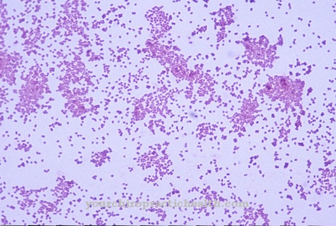
The Microsporum canis is a parasite on the skin of cats and dogs. The fungus spreads to humans or other animals through zoonosis. In southern countries almost all stray animals are infected with the pathogen. The fungus forms cotton-wool-like and limited colonies on nutrient media, which appear creamy-white to orange-yellow. In the macroscopic picture it has septate hyphae and smooth club-like microconidia. The individual macroconidia have a spindle shape and are up to 25 by 110 micrometers in size. They each have up to 18 chambers, have knotty ends and rough walls.
Microsporum gallinae is also a parasitic skin fungus that often causes dermatophytosis, especially in birds. As a zoonotic pathogen, it can also cause cross-species infections. This fungus forms slightly fluffy, velvety white colonies and, under the microscope, shows septate hyphae with round to pear-shaped microconidia up to eight by 50 micrometers in size. The microconidia show a slight curvature and are equipped with fine spines at the ends.
Another representative of the microsporum is the skin parasite Microsporum gypseum. It is predominantly geophilic and is transmitted through the ground. In humans, transmission leads to the image of the gardener microsporia, but horses and cats can also be carriers of the pathogen due to the zoonosis. The fungus forms fluffy white colonies with septate hyphae and club-shaped microconidia up to 16 by 50 micrometers in size. The symmetrically arranged, rough and thin-walled microconidia are rounded at the ends.
Humans become infected with microsporum mainly when they come into contact with contaminated animals, more rarely when they come into contact with the ground. A smear infection from person to person is also possible.
Fungi of the species reproduce purely vegetatively or by spores. These so-called conidia are formed in an asexual way. As they grow, they gain energy from the breakdown of carbohydrates and keratin, which they do with the help of the enzyme keratinase.
Illnesses & ailments
The microsporum is clinically pathogenic and is considered to be the causative agent of microspories. These dermatophytoses of the skin manifest themselves in the form of skin mycosis. Tinea corporis is characterized by red scaly efflorescences that start centrally and gradually spread to the periphery as the infection progresses. In addition, fungi of the species Microsporum often cause hair mycosis. This tinea capitis is mainly associated with the microsporum canis and makes the hair brittle.
Animals in particular, but also humans, can be silent carriers of the infection. If so, they will not have any symptoms, but they can nonetheless pass the fungus on. Depending on the region of the infestation, the doctor takes examination material from the edge of the lesion or from hair for the purpose of diagnosis. Pathogens are detected microscopically or in culture, for example on Sabouraud agar.
For local therapy of the infection, patients are prescribed different drugs. Fluconazole and itraconazole are particularly promising active ingredients in the treatment of various fungal diseases of the skin and hair. Voriconazole also helps against dermatophytes in particular.
Alternatively or in combination, active ingredients such as terbinafine or triazoles can be used. As a rule, however, this therapeutic step only takes place in the event of extremely severe infestation. The doctor prescribes griseofulvin even more rarely, which was used far more often in the treatment of fungal diseases.
People are particularly often infected with the pathogens while on vacation in southern regions. This connection is mainly due to the high rate of the infestation of the strays there.


.jpg)


.jpg)




.jpg)

