The heart consists of a right and a left half and is divided into four chambers. The heart septum, also known as the septum cordis, runs lengthways between the two halves of the heart. The septum separates the four Ventricles into a left and right atrium, and a left and right ventricle. The terms also become synonymous Cardiac ventricle or Ventriculus cordis used.
What is the heart chamber?
The left ventricle is part of the body's circulation and is connected to the left atrium. It is responsible for supplying the body's circulation via the aorta with the blood freshly arriving from the lungs. The right ventricle is part of the pulmonary circulation and is connected to the right atrium.
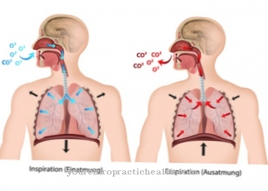
It pumps the venous blood, which has absorbed large amounts of carbon dioxide as a breakdown product from the cells, into the pulmonary vessels. There the degradation product is exhaled and the blood can take up oxygen again. The arterial blood then flows through the left ventricle into the body's circulation.
Anatomy & structure
The fist-sized heart is embedded between the two lungs. It is located above the diaphragm. The heart wall has three layers. The endocardium forms the inner lining of the heart, the myocardium (heart muscle) a large part of the heart wall. The epicardium covers the coronary vessels and the surface of the heart.
It is made very thin and regularly releases a clear liquid so that the heart glides in the pericardium during the pumping process. The pericardium consists of connective tissue that surrounds the heart. It consists of the left and right halves and is divided into four chambers. The two halves of the heart are separated longitudinally by the septum (heart septum). This divides the four chambers into a right and a left heart chamber as well as a right and a left atrium. Chambers and atria are horizontally separated from each other by so-called sail valves.
The right valve is called the tricuspid valve and the left is called the mitral valve. These heart valves work on the principle of a check valve. They ensure that the blood flow within the heart is only one direction. The right half of the heart is facing the anterior chest wall (ventral), while the left half of the heart is facing back (dorsal). The left ventricle is part of the body's circulation, while the right ventricle is part of the pulmonary circulation.
Function & tasks
The heart connects the pulmonary and body circulation. According to its anatomy, it constantly pumps blood through the body and supplies the organs with oxygen. A healthy heart beats about 70 times per minute and carries 70 milliliters of blood with each heartbeat, which corresponds to a blood volume of five liters per minute.
A complicated system of excitation conductors ensures that the pumping function runs smoothly. The sinus node located in the right atrium generates the electrical impulse necessary to contract the heart muscle. From here, the electrical impulses travel along the atria and ventricles and spread to the top of the heart. The inferior and superior vena cava flow into the right atrium. The venous (low-oxygen) blood from the body's circulation flows through these vena cava to the heart. The blood then flows from the right atrium into the right ventricle and via the pulmonary artery (pulmonary artery) into the lungs.
The pocket-shaped pulmonary valve is located between the heart and pulmonary artery. Arterial, oxygen-saturated blood flows from the lungs into the left atrium via the pulmonary veins. It is then passed on to the left ventricle and returns to the organs via the aorta (main artery). At the point of origin of the aorta there is also a pocket valve, the aortic valve. The heart is supplied from outside by means of small blood vessels. These blood vessels are called coronary arteries or coronary arteries. They originate from the main artery branching off from the left ventricle.
The right and left coronary arteries form the coronary vessels. They have numerous fine branches. Their job is to regularly supply the heart muscle with oxygen. The heart's pumping process takes place regularly in three steps. The first step is the filling phase (diastole). The heart muscle relaxes. Oxygen-deficient blood flows through the vena cava into the right atrium and then into the right ventricle. At the same time, oxygen-saturated blood flows from the lungs to the left atrium. It is then passed on to the left ventricle. The leaflet valves close when the chambers have a higher filling pressure than the atria.
The second step is the tension phase. The two atria contract and increase the amount of blood in the chambers. The third step is the expulsion phase (systole). The heart muscle contracts and the blood in the chambers flows through the large blood vessels into the body and lungs. The closed leaflet valves prevent the blood from flowing back into the atria. As the evacuation increases, the pressure in the heart chambers decreases.
The tightly closed pocket flaps prevent the blood from flowing back from the large vessels into the heart chambers. The drop in pressure causes the chambers to refill with the blood in the atria. Now the cycle repeats itself with diastole and systole.
Diseases
In left heart failure, the left ventricle no longer works adequately due to a pump weakness. Difficulty breathing occurs and breathing is usually accelerated (tachypnea). Patients experience cold sweats, coughs and a rattle in their lungs.
Other complaints are pulmonary congestion, pulmonary edema and feelings of restlessness. The medical term is asthma cardiale. If a patient has right heart failure, water builds up in the ankles and shins. Sick people feel an increased urge to urinate, as water from the tissue washes out into the blood and is excreted with the urine. Skin edema occurs in the area of the genitals, buttocks and flanks. Because the blood builds up in the veins in front of the right heart, the neck veins are very full.
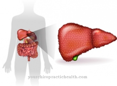
The venous blood is deposited in various organs; the liver can enlarge (congested liver) and water can accumulate in the abdomen (ascites). An inflammation is possible in the gastric veins, which causes inflammation of the gastric mucosa (congestive gastritis). It is accompanied by a feeling of fullness and loss of appetite. Only in rare cases do these two heart diseases occur separately. Most patients suffer from global heart failure, in which both heart chambers no longer work properly.





.jpg)

.jpg)
.jpg)
